
Fig. 4
- ID
- ZDB-FIG-041230-4
- Publication
- Kapsimali et al., 2004 - Inhibition of Wnt/Axin/{beta}-catenin pathway activity promotes ventral CNS midline tissue to adopt hypothalamic rather than floorplate identity
- Other Figures
- All Figure Page
- Back to All Figure Page
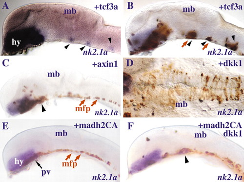
Suppression of Wnt signalling in combination with activation of Nodal signalling promotes hypothalamic marker gene expression. Lateral (A,B,C,E,F) and ventral (D) views of brains of embryos at 24-somite stage with anterior to the left in which cells (brown) overexpressing hdl/tcf3a (A,B), axin1 (C), dkk1 (D), madh2CA (E) or madh2CA+dkk1 (F) were transplanted into the prospective hypothalamus/anterior floorplate at 60-65% epiboly. A and B show the same embryo before and after immunohistochemistry for Gfp. In A,B,C,F, black arrowheads point to ectopic nk2.1a expression in the medial floorplate and/or adjacent cells. In B,C,E, red arrows point to cells that integrated in the medial floorplate. In D, Dkk1+ cells (brown) mainly integrate in more lateral positions in the neuroepithelium compared to the Axin1+ and Madh2CA+ cells that integrate in the medial floorplate (brown, red arrows) of the embryos in C and E respectively. Abbreviations: hy, hypothalamus; mb, midbrain; mfp, medial floorplate; pv, posterior-ventral hypothalamus. |