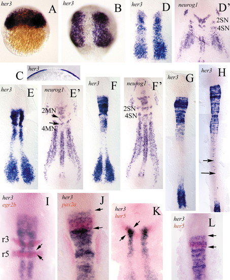
Distribution of her3 transcripts revealed by whole-mount in situ hybridisation. (A) 30% epiboly. her3 transcription is first detected in the blastoderm. (B) 80% epiboly. The single initial transcription domain has split into two, each of which lies within the presumptive neural primordium. (C) Cross-section of B showing transcription in the epiblast. (D) Flat preparation of a tailbud-stage embryo. The her3 expression domains form two broad longitudinal stripes in the intermediate lateral regions of the neural plate. Compare this with the embryo in D', which was hybridised with a neurog1 probe (see Blader et al., 1997; Geling et al., 2003). The two transcription patterns are complementary. E,E' (two somites) and F,F' (four somites) show that the her3 domains occupy the regions in which neurog1 is not transcribed. 2MN and 4MN, motoneurones of rhombomeres 2 and 4. 2SN and 4SN, sensory neurones of rhombomeres 2 and 4. (G,H) Eight-somite (G) and 16-somite stage (H) show that transcripts of her3 later become undetectable in anterior regions but continue to be expressed in single cells within caudal regions of the developing spinal cord (arrows). (I,J) Double in situ hybridisation with her3 (blue) and egr2b (red), and with her3 (blue) and pax2a (red), respectively. Note that the her3 domain progressively extends into rhombomere 5 (r5) (arrows). (K,L) In situ hybridisation with her3 (blue) and her5 (red), to show that the rostral margin of the her3 domain corresponds to the anterior border of the midbrain primordium. The her5 domain is included within the her3 domain (between arrows). Refer to the text for further details. Anterior is towards the top in all embryos.
|
