
- Title
-
Homologs of the Xenopus developmental gene DG42 are present in zebrafish and mouse and are involved in the synthesis of Nod-like chitin oligosaccharides during early embryogenesis
- Authors
- Semino, C.E., Specht, C.A., Raimondi, A., and Robbins, P.W.
- Source
- Full text @ Proc. Natl. Acad. Sci. USA
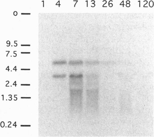
Northern blot hybridization. Zebrafish total RNA was extracted from embryos at the times indicated by the top row (hours after fertilization). The probe was DNA of the cloned PCR fragment of the zebrafish DG42 homolog. The origin (o) and size standards (in kilobases) are designated by bars. |
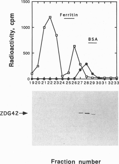
Distribution of the zebrafish DG42-like protein in a size exclusion Superose 6 column and fractionation of hyaluronate and chitin oligosaccharide synthase activity. A sample of 300 zebrafish embryos from late gastrula stage were lysed in 200 μl of 1% digitonin buffer and injected into a Superose 6 column equilibrated with 0.1% digitonin buffer as described in Materials and Methods. Fractions 19-33 were used to detect activity of hyaluronate synthase (□) (using labeled UDP-[14C]GlcA and unlabeled UDP-GlcNAc) or chitin oligosaccharide synthase activity (●) (using labeled UDP-[3H]GlcNAc and unlabeled UDP-GlcA) (see Material and Methods). The same methodology used in Table 1 for analysis of HA and chitin oligosaccharides was used here. An aliquot (100 μl) of each fraction was concentrated, resuspended in 1% SDS loading buffer, boiled for 2 min, and applied to a 10% SDS-polyacrylamide gel. After electrophoresis, proteins were transferred to a nitrocellulose membrane and used to develop a Western blot with the anti-DG42 antibody as described in Materials and Methods. The figure at the bottom indicates with an arrow the fractions where zebrafish DG42-like protein was detected (ZDG42). Superose 6 has an exclusion size of 5 x 10³ kDa. Blue dextran (2 x 10³ kDa), ferritin (440 kDa), and bovine serum albumin (66 kDa) were used to calibrate the column. |