FIGURE SUMMARY
Title

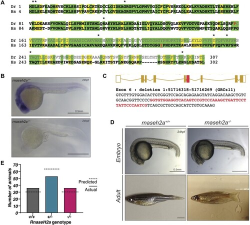
Second generation lethality in RNAseH2a knockout zebrafish

Authors
Thomas, R.C., Zaksauskaite, R., Al-Kandari, N.Y., Hyde, A.C., Abugable, A.A., El-Khamisy, S.F., van Eeden, F.J.
Source
Full text @ Nucleic Acids Res.

Generation of rnaseh2a−/− knockout zebrafish and initial characterization. (A) Protein alignment between zebrafish, Danio rerio (Dr) and Homo sapiens (Hs) RNaseH2a. In the zebrafish protein, the amino acids that are lost as a result of the mutation are greyed out, the last homologous amino acid is highlighted red. Asterisk show sites of human AGS mutations, a critical tyrosine required highlighted in red. (B) WISH staining shows universal RNaseH2a expression in wild-type zebrafish embryos at 24hpf, lower image shows sense probe as control. (C) In exon 6 (red), a 49bp deletion was created utilizing the CRISPR/Cas9 system. Exon 6 sequence is shown below with deleted bases in red. The deletion results in a premature stop codon and truncated protein of 205aa in length. (D) Embryos resulting from an rnaseh2a+/− incross do not show any visible phenotype and are able to develop to adulthood with no external phenotype. NB. spotty pigment is due to a background mutation in laboratory zebrafish. (E) Inheritance of the rnaseh2a mutation is in a homozygous recessive manner at 24hpf and displays mendelian inheritance. X2 = 2.66, with two degrees of freedom; two-tailed P value of 0.529.

Offspring of rnaseh2a−/− adults have severe developmental defects, increased DNA damage, increased ribonucleotides and an upregulated inflammatory response. (A) Acridine Orange staining of apoptotic cells (green) in 24hpf embryos from homozygous and wild-type incrosses. (B) Quantification of acridine orange positive cells from (A). Unpaired t-test, ****P <0.0001, ±SD (n = 12, single embryo). (C) Schematic of single ribonucleotide cleavage assay. (D) Whole embryo lysate from 24hpf embryos were incubated with dsDNA substrate containing a single ribonucleotide. Positive and negative controls were purified RNaseH2 and lysis buffer respectively. DNA was tagged with Cy5. (E) Quantification of cleavage (%) in activity assay, ±SD, one-way ANOVA, P <0.05 (n = 3, 20 pooled embryos). (F) Gel of alkaline (NaOH) and control (NaCl) treated DNA of 24hpf embryos from homozygous and wild-type crosses. (G) Densitometry plot of DNA intensity produced with R. (H) Image quantification showing ratio of long and short fragments. Student's t-test **P< 0.01, ± SEM, n = 3. (I) γH2AX staining (green) in tails of 24hpf rnaseh2a−/− embryos. Nuclei stained with DAPI (blue). (J) Quantification of γH2AX foci per nucleus in tail tip of individual embryos (>100 nuclei per embryo, 3 or 4 per genotype shown). Red lines show average per embryo and SEM. Unpaired t-test, Welch's correction, ***P <0.001, n ≥ 3). (K) qPCR of interferon stimulated genes (ISG15, mxa, IFNΦ), proinflammatory response markers (TNFa, ILb, IL6) and senescence markers (p21). ****P <0.0001, ***P <0.001, **P <0.01, ±SD (n = 3, 20 pooled embryos).

rnaseh2a−/− adult fish have a high incorporation of ribonucleotides but no increased inflammatory response. (A) RNaseH2 activity assay on brains from adult fish. (B) Activity assay of 5dpf embryos. (C) Quantification of cleavage assay on brains, ±SD, one way ANOVA, P <0.05, n = 3. (D) Quantification of cleavage activity of d5 embryos, ±SD, one way ANOVA, P <0.05, n = 3. (E) Alkaline treatment of DNA from brains from adults shows an increase of ribonucleotides in homozygote adults, as shown by smearing on a gel. (F) Quantification of alkaline treatment of adult brains. Image analysis shows that the ratio of long to short DNA fragments is significantly reduced in adult rnaseh2a−/− brains. Student's t-test, n = 3 ± SEM, *P< 0.05. (G) Alkaline treatment of DNA from on 5dpf embryos showing no increase in DNA smearing in homozygous embryos. (H) Quantification of alkaline treatment of 5dpf embryos. Student's t-test, n = 3 ± SEM. (I) γH2AX staining 5dpf embryo tails from a heterozygous incross. Quantification of foci per nuclei shows no significant difference between rnaseh2a−/− and rnaseh2a+/+ siblings. Student's t-test, n = 3, P <0.05. (J) Western blot for γH2AX in brain samples from individual adult zebrafish. Quantification showed no significant difference between rnaseh2a−/− and rnaseh2a+/+ siblings. Student's t-test, ±SD, P <0.05. n = 3. (K) Expression of interferon stimulated genes (ISG15, mxa and IFNphi), inflammatory response genes (TNFα, IL1β and IL6) and senescence markers (p21) were measured in tissues isolated from rnaseh2a-/- 19 month adults and in 5dpf rnaseh2a-/- embryos from a heterozygous incross. Multiple unpaired t-tests with Welch correction, *P <0.05, **P <0.01. ***P <0.001, ****P <0.0001, ±SD (n = 3).

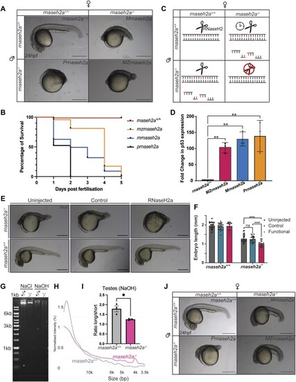
Re-introduction of RNaseH2a is unable to rescue the developmental phenotype. (A) Embryos resulting from various crosses with homozygous rnaseh2a−/− parents. (B) Survival curve of embryos with various homozygous parents (n = 110). (C) Schematic of predicted mechanism of detrimental phenotypes resulting from each parental cross. (D) RTqPCR analysis of a DNA damage sensitive p53 reporter (Δ113p53) shows significant upregulation of expression in all embryos resulting from a homozygote parent. ±SD, one-way ANOVA, **P <0.001 (n = 3, 20 pooled embryos). (E) Microinjection of RNaseH2a mRNA caused a more severe phenotype in embryos from a rnaseh2a−/− incross compared with the control or uninjected embryos. Embryos from a wild-type incross were uninjected. (F) Quantification of embryo size, ±SD, two-way ANOVA, ****P <0.0001 (n = 16). (G) Gel analysis of alkaline treated DNA from testes of adult males show an increase in ribonucleotides in rnaseh2a-/- adult males. (H) Density plot of alkaline treatment of testes from adult zebrafish. (I) Ratio of long to short DNA fragments is significantly reduced in rnaseh2a−/− testes DNA after alkaline treatment. Student's t-test, n = 3 ± SEM, *P< 0.05. (J) Microinjection of a p53 morpholino visibly improves the development of all embryos with one or two homozygous parents (injected embryos are from same batch as the embryos shown panel A).

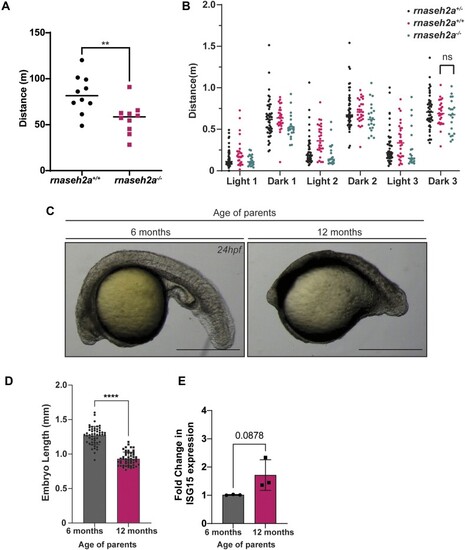
Embryos from older rnaseh2a−/− fish are significantly underdeveloped compared with embryos from younger fish. (A) Movement analysis of adult zebrafish revealed the distance moved by rnaseh2a−/− is significantly shorter than rnaseh2a+/+ adults. Unpaired t-test, n = 10, individual adults, ±SD, ** P < 0.001. (B) Photomoter response of embryos from rnaseh2a+/− parents show no significant difference in distance moved by rnaseh2a+/+ compared with rnaseh2a−/− ±SD, unpaired t-test, n = P < 0.05. (C) Representative images of embryos from 6 month old and 12 month old rnaseh2a−/− zebrafish. (D) Quantification of embryo length from old and young zebrafish n = 36, individual embryos. Unpaired t-test **** P < 0.0001. (E) RTqPCR showing an increase in the expression level of ISG15 in embryos resulting from older rnase2a−/− adults. n = 3, 20 pooled embryos. Unpaired t-test P < 0.05.

Acknowledgments
This image is the copyrighted work of the attributed author or publisher, and ZFIN has permission only to display this image to its users. Additional permissions should be obtained from the applicable author or publisher of the image. Full text @ Nucleic Acids Res.