
- Title
-
Hhex and Prox1a synergistically dictate the hepatoblast to hepatocyte differentiation in zebrafish
- Authors
- Jin, Q., Hu, Y., Gao, Y., Zheng, J., Chen, J., Gao, C., Peng, J.
- Source
- Full text @ Biochem. Biophys. Res. Commun.

Early endoderm patterning is not obviously affected in the hhex and prox1a double mutant. A-C. WISH using foxa3, gata4, and gata6 probes on the progenies derived from the intercrosses of the male and female hhex+/− prox1a+/− double heterozygotes at 34hpf (A), 2dpf (B) and 5dpf (C). The number of total embryos genotyped (denominator) and the number of embryos exhibiting the displayed phenotype (numerator) are provided at the bottom right. Yellow outline, the foregut region giving rise to the prospective liver, pancreas and hepatopancreatic duct region. in, intestinal bulb; lv, liver primordium; pc, pancreatic bud. Scale bar, 200 μm. (For interpretation of the references to color in this figure legend, the reader is referred to the Web version of this article.) |
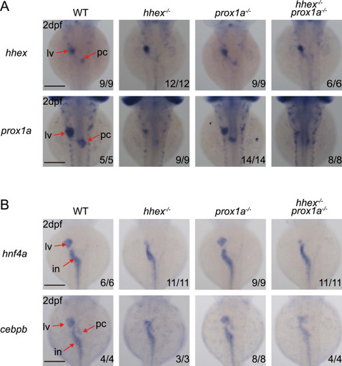
The prospective hepatic region is successfully specified in the hhex and prox1a double mutant. A and B. WISH using the hhex and prox1a probes (A) and hnf4α and cebpb probes (B) on the progenies derived from the intercrosses of the male and female hhex+/− prox1a+/− double heterozygotes at 2dpf. The number of total embryos genotyped (denominator) and the number of embryos exhibiting the displayed phenotype (numerator) are provided at the bottom right. in, intestinal bulb; lv, liver primordium; pc, pancreatic bud. Scale bar, 200 μm. |
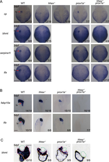
hhex and prox1a double mutant lacks a detectable expression of molecular markers at the prospective liver site. A. WISH analysis of the molecular marker cp, bhmt, sperpina1l, and tfa in the embryos obtained from the intercrosses of male and female hhex+/− prox1a+/− double heterozygotes at 2dpf. Scale bar, 200 μm. B. WISH analysis of the liver molecular markers fabp10a and tfa in 5dpf zebrafish embryos obtained from the intercrosses of male and female hhex+/− prox1a+/− double heterozygotes. Scale bar, 200 μm. C. The bhmt post-WISH embryos were sectioned to facilitate the visualization of its expression in the liver. Green outline, intestine; yellow outline, YSL; red outlined, liver. Scale bar, 100 μm. In A-C, the number of total embryos genotyped (denominator) and the number of embryos exhibiting the displayed phenotype (numerator) are provided at the bottom right. lv, liver. (For interpretation of the references to color in this figure legend, the reader is referred to the Web version of this article.) |

Bmp2a failed to induce a hepatic fate in the hhex and prox1a double mutant. A. WISH using cp and fabp10a two probes on the embryos obtained from the intercrosses of male and female hhex+/− prox1a+/− double heterozygotes at 3dpf after the injection of the bmp2a mRNA at one-cell stage. The number of total embryos genotyped (denominator) and the number of embryos exhibiting the displayed phenotype (numerator) are provided at the bottom right. lv, liver. Scale bar, 200 μm. B. A model depicts the roles of Hhex and Prox1a in dictating the potency of the prospective hepatoblasts to be differentiated into hepatocytes and cholangiocytes. Left, Hhex and Prox1a protects the hepatic fate by transcriptionally inhibiting cdx1b and pdx1. Right, Loss-of-function of hhex leads to intestinalized extrahepatic duct while the prox1a−/− mutant displays an intestinalized liver due to ectopic expression of cdx1b. Combinatory loss-of-function of Hhex and Prox1a leads to a liver less phenotype. Pink color, hepatocytes; green color, cdx1b-positive domain. (For interpretation of the references to color in this figure legend, the reader is referred to the Web version of this article.) |