
- Title
-
Loss of a cilia-associated gene, pkd1l1, causes biliary defects in zebrafish: Implications for Biliary Atresia Splenic Malformation
- Authors
- Ali, R.Q., Meyer-Miner, A., David-Rachel, M., Lee, F.J.H., Wilkins, B.J., Karpen, S.J., Ciruna, B., Ghanekar, A., Kamath, B.M.
- Source
- Full text @ Dis. Model. Mech.
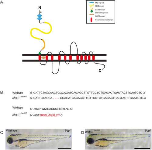
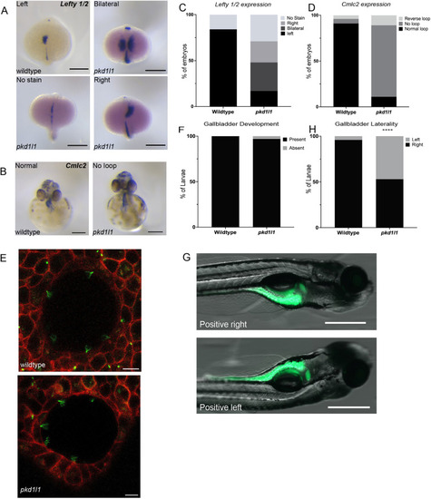
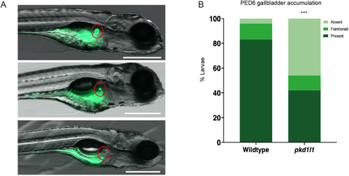
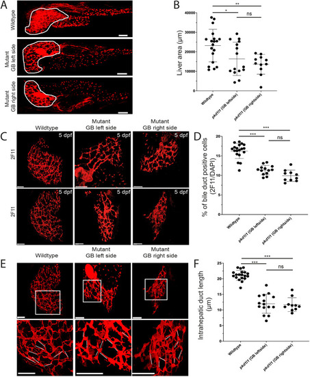
PHENOTYPE:
|
EXPRESSION / LABELING:
PHENOTYPE:
|
PHENOTYPE:
|
PHENOTYPE:
|
PHENOTYPE:
|