
- Title
-
A novel in-frame deletion in MYOT causes an early adult onset distal myopathy
- Authors
- Guglielmi, V., Pancheri, E., Cannone, E., Nigro, V., Malatesta, M., Vettori, A., Giorgetti, A., Torella, A., Aurino, S., Cisterna, B., Marchetto, G., Tomelleri, G., Tonin, P., Schiavone, M., Vattemi, G.
- Source
- Full text @ Clin. Genet.

(A) Pedigree of the family. Arrow indicates the proband. Solid black symbols denote affected family member. (B) Sanger. UCSC graphic view of the N-terminal region of MYOT (upper panel). Electropherogram showing the heterozygous Tyr4_His9del in-frame deletion in exon 2 of MYOT (Lower panel). (C) Model of the human myotilin. The atom indexes are colored from red to blue depending on their sequence position. In green the p.Tyr4_His9del residues. The zoomed region illustrates the interaction between the residues involved in the deletion. The red circle shows the salt bridge formed by Arg6 and Glu5. [Colour figure can be viewed at wileyonlinelibrary.com] |

(A, B) Morphological features observed at the light microscope and quantitative analysis of the phenotypes of 24 hpf embryos injected with human wtMYO or mutMYO mRNA. (C) Representative lateral views of a single confocal projection (z-stack) of mutMYO, wtMYO, and control embryos at 48 hpf stained with phalloidin. Skeletal muscle fibers appear less consistent and compact in mutMYO-injected embryos compared to wtMYO-injected or not injected (control) larvae. (D) Transmission electron micrographs of skeletal muscles from embryos injected with wtMYO mRNA (a, b) and mutMYO mRNA (c, d). Note the irregular arrangement and the heterogeneous thickness of myofibres in mutant embryos (c) compared to controls (a). High magnification images (b, d) show in mutant embryos well-organized Z lines but I-bands wider than in controls. Bars: 1 μm (a, c); 200 nm (b, d). [Colour figure can be viewed at wileyonlinelibrary.com] |
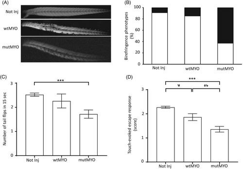
(A, B) Birefringence analysis of wildtype embryos injected with Tol2 plasmids harboring either human wild-type MYOT full cDNA (wtMYO) or human MYOT c.11_28del (mutMYO) was performed at 48 hpf. In (A), representative lateral images of single non-injected, wtMYO-, and mutMYO-injected embryos. Bar: 50 μm. The bar graph in (B) reports the birefringence phenotypes observed in not injected (n = 200), wtMYO (n = 55), and mutMYO (n = 76) embryos. Data are plotted as total percentage of fish showing wildtype (white closed bars) or myopathic (black closed bars) birefringence phenotypes. (C) Number of spontaneous coiling events (tail flips) performed in 15 s have been recorded at 24 hpf in non-injected (n = 330), wtMYO-injected (n = 80) and mutMYO-injected (n = 85) embryos and plotted in the bar graph. Values are reported as the average of the number of tail flips recorded for single embryos at 24 hpf ± SEM ***p < 0.001. (D) Touch-evoked escape response at 48 hpf was checked in non-injected (n = 342), wtMYO (n = 86), and mutMYO (n = 78). Arbitrary values (0–3) have been assigned to each larva based on the observed response. Data in the bar graph are reported as the average of escape values for single embryos ± SEM *p < 0.05, **p < 0.01, ***p < 0.001. |