FIGURE SUMMARY
Title

The translation initiation factor homolog eif4e1c regulates cardiomyocyte metabolism and proliferation during heart regeneration

Authors
Rao, A., Lyu, B., Jahan, I., Lubertozzi, A., Zhou, G., Tedeschi, F., Jankowsky, E., Kang, J., Carstens, B., Poss, K.D., Baskin, K., Goldman, J.A.
Source
Full text @ Development

Eif4e1c is both unique to and shared by all aquatic vertebrates. Shown is a phylogeny of eIF4E1 orthologs from a sampling of the species considered (see Fig. S1 for full analysis). Eif4e1c is ancestral to the canonical eIF4E1A split from its variant, eIF4E1B. Terrestrial species have a canonical and eIF4E1B ortholog (pink and purple). All aquatic species have a canonical variant: eight species with a duplication (blue and green), two species retain only one variant of the duplication, and the Scyliorhinus canicula (small-spotted catshark) canonical clusters with terrestrial eIF4E1A. All 12 aquatic vertebrate retain an Eif4e1c family member. Shown are images of each of the aquatic vertebrate species to highlight the diversity considered. Only five of 12 aquatic vertebrates shown here retain an eIF4E1B variant. Estimates are made using maximum likelihood and the IQ-TREE. Nodes with bootstrap support <0.85 are marked with their respective values, all other nodes had support values of 0.85 or higher.

Highly conserved Eif4e1c-specific amino acids form a novel patch along the surface of the protein. (A) ClustalW sequence alignment of human versus fish Eif4E orthologs. Highlighted are universally conserved amino acids required for cap binding (green) and eIF4G recruitment (pink). Residues that distinguish EIF4E1A family members (gray) from EIF4E1B family members (black) are also highlighted. The six amino acids most responsible for strong cap binding are marked by red carets. The Eif4e1c family conserved residues are highlighted yellow with red letters. The seven residues that are identical in all EIF4E1A and EIF4E1B family orthologs throughout evolution but are uniquely changed in Eif4e1c family members are highlighted in red. Amino acids that diverge between mouse and human are bolded in the human sequence. (B) PyMOL rendition of zebrafish Eif4e1c mapped along the human structure of EIF4E1A bound to a methylated cap (green) and an EIF4G peptide (orange). The tryptophans required for both associations are conserved and highlighted in yellow. All Eif4e1c-specific residues are highlighted in red. (C) The abundance of each of the transcripts shown in the key were derived from RNAseq datasets from different adult tissues. Expression is determined relative to mob4. The FPKM for the three orthologs were similar in the fin. Values shown have been normalized to expression levels in the fin. Where error bars are presented, at least three biological replicates were used to determine the average and s.d. Only testis, ovary and oocyte had two replicates. Dots show individual data points. SC, spinal cord. (D) Time course of RNAseq of different developmental stages in embryos. Normalized sequencing reads for the four different EIF4E1 orthologs are shown.

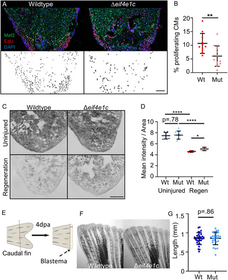
Δeif4e1c mutants have impaired growth and poor survival. (A) Schematic of the CRISPR-mediated deletion of eif4e1c. Red rectangles represent coding exons, and the white box represents exons with untranslated regions. (B) Fish were grown together, genotyped at the indicated stage and immediately measured from jaw to the caudal fin bifurcation (3-month average wild type=1.82 cm, mutant=1.62 cm; Welch's t-test, P=0.0156; n=24 and 23, respectively; 6-month average wild type=2.48 cm, mutant=2.37 cm; Mann–Whitney, P=0.121; n=20 and 20, respectively). (C) After measuring length, fish were dried off as much as possible and weighed. The 8 wpf data (gray box) is shown on the right with a different scale for the y-axis. [8-week (blue) average wild type=35.75 mg, mutant=22.82 mg; Welch's t-test, *P=0.0392; n=24; 3-month (green) average wild type=146.5 mg, mutant=103.1 mg; Welch's t-test, **P=0.0014; n=24 and 23, respectively; 6-month average (red) wild type=344.6 mg, mutant=261.7 mg; Welch's t-test, ***P=0.0002; n=20). (D) Uninjured heart ventricles from wild-type and mutant fish were sectioned and stained for muscle using antibodies to myosin heavy chain (MHC) and the cardiac transcription factor Mef2c to identify CM nuclei. Scale bar: 100 µm. Note that adjacent hearts have been removed from images for presentation purposes. (E) Numbers of Mef2c-positive cells were counted with MIPAR. The 8 wpf data (red box) is shown on the right with a different scale for the y-axis. [8-week (blue) average wild type=1078, mutant=848; Welch's t-test, *P=0.0383, n=17; 3-month (green) average wild type=6061, mutant=4557; Welch's t-test, **P=0.0052, n=10 and 8, respectively). (F) Left: Western blot of whole-cell extracts from zebrafish hearts using an antibody directed towards canonical Eif4ea (top) and Tnnt2a (bottom). Right: Quantification of western blots with total Eif4ea levels normalized to the sarcomeric protein Tnnt2a as a measure of total cardiac mass (average increase=1.82; Welch's t-test, *P=0.0334, n=4). (G) Immunofluorescence of canonical Eif4ea/b (top, grayscale; bottom, green) and MHC (blue, bottom). Scale bar: 100 µm. For each panel, the lighter shade color is used for mutants, individual data points are represented by dots, and error bars represent mean±s.e.m. Mut, mutant; Wt, wild type.

Ribosome profiling indicates translational changes in eif4e1c mutant hearts. (A) Example of fluorescence in the hearts of OPP-injected fish. Scale bar: 100 µm. (B) Quantification of OPP fluorescence shows no significant difference between mutant and wild-type fish (normalized fluorescence average wild type=140,279 arbitrary density units (adu)/µm2, mutant=145,096 adu/µm2; Mann–Whitney, P=0.806; n=15). Error bars represent mean±s.e.m. (C) Diagram of the ribosome profiling method. Ribosome-bound mRNA is purified from wild-type and mutant hearts and then subjected to micrococcal nuclease (MNase) digestion. Fragments of mRNA protected by being bound by ribosomes are then purified and subjected to high-throughput sequencing. (D) Differences (mutant/wild type) in mRNA abundance (log2) are plotted versus changes in the abundance of ribosome-protected fragments (RPF). Genes for which translation does not change significantly are plotted in gray, genes for which translation is decreasing in the mutant are plotted in red and genes for which translation is increasing in the mutant are plotted in blue. Decreasing genes mentioned in the text are colored gold and increasing genes mentioned in the text are colored green. (E) High-throughput sequencing browser tracks for two genes (blue arrows) that are involved in cell-cycle progression (cdc123 and mapre1a). Wild-type (WT) data is shown in blue, and mutant (MUT) data is shown in green. The top two tracks (mRNA) show the abundance of mRNA (as measured from RNAseq) and the bottom two tracks show the abundance of RPFs. Dashed red boxes highlight decreasing levels of RPF where mRNA levels are comparable. (F) Time course of oxygen consumption rates (OCR) from mitochondria measured by the Seahorse analyzer. Dotted lines indicate time points of drug injection. Readings were normalized to the number of mitochondria using qPCR. The first injection (I1) is of ADP to stimulate respiration, the second injection (I2) is of oligomycin to inhibit ATP synthase (complex V) decreasing electron flow through transport chain, the third injection (I3) is of FCCP to uncouple the proton gradient, and the fourth injection (I4) is of antimycin A to inhibit complex III to shut down mitochondrial respiration. Error bars represent mean±s.e.m. (G) Basal respiration is calculated as the OCR average after addition of ADP (I1 to I2) subtracting the non-mitochondrial respiration after injection of antimycin A (after I4) (mean wild type=60.01, mutant=42.25; Welch's t-test, ****P<0.0001; n=15). Maximal respiration is calculated as the OCR average after FCCP addition (I3 to I4) subtracting the non-mitochondrial respiration after injection of antimycin A (after I4) (mean wild type=68.73, mutant=52.19; Welch's t-test, ****P<0.0001; n=15). Error bars represent mean±s.e.m. Mut, mutant; Wt, wild type.

Deletion of eif4e1c impairs CM proliferation in regenerating hearts. (A) Images of sectioned amputated ventricles (7 dpa) from wild-type and Δeif4e1c mutant fish. Sections are stained for Mef2c (green) and EdU (red). Double-positive cells are highlighted in black below using a MIPAR software rendition. Scale bar: 100 µm. (B) Quantification of CM proliferation indices (Mef2/EdU double-positive over total Mef2 positive) in 7 dpa ventricles (average wild type=10.68%, mutant 6.02%; Welch's t-test, **P=0.0054; n=11 and 13, respectively). Error bars represent mean±s.e.m. (C) Cryosections of fresh mutant and wild-type hearts were stained for Succinate dehydrogenase activity. Shown are uninjured hearts (top) and hearts that were ablated genetically using Z-CAT (7 days-post-incubation) (bottom). Scale bar: 20 µm. (D) The mean signal intensity was calculated per given area for Sdh-stained hearts (uninjured average wild type=7.47, mutant=7.57; n=6 and 8, respectively; regeneration average wild type=4.59, mutant=5.06; n=4). Welch's t-test was used to calculate significance (wild type versus mutant uninjured=0.781, wild type uninjured versus wild type regeneration ****P<0.0001, mutant uninjured versus mutant regeneration ****P<0.0001). For every replicate (n=4) the mutant Sdh activity during regeneration (pink) was higher than the wild-type activity during regeneration (red). Error bars represent mean±s.e.m. (E) Diagram of caudal fin regeneration experiments. The blastema is the highly proliferative zone of growth that forms the new fin. (F) Caudal fins were amputated ∼50% in Δeif4e1c mutants and their wild-type siblings and shown are images of fin regrowth 4 days later. (G) There was no significant difference between fin growth in wild type (blue) and mutant (light blue) fish (average wild type=0.849 mm, mutant 0.857 mm; Welch's t-test, P=0.861; n=30 and 26, respectively). Error bars represent mean±s.e.m. Mut, mutant; Wt, wild type.

Acknowledgments
This image is the copyrighted work of the attributed author or publisher, and ZFIN has permission only to display this image to its users. Additional permissions should be obtained from the applicable author or publisher of the image. Full text @ Development