FIGURE SUMMARY
Title

In vivo Drug Screening to Identify Anti-metastatic Drugs in Twist1a-ERT2 Transgenic Zebrafish

Authors
Nakayama, J., Makinoshima, H., Gong, Z.
Source
Full text @ Bio Protoc

Twist1a-ERT2/xmrk double transgenic zebrafish offers an in vivo drug screening platform for the identification of anti-metastatic drugs.

A. Representative images of the dissemination of mCherry-labeled hepatic cells from the liver in
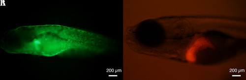
 Twist1a-ERT2/xmrk double transgenic zebrafish at 16 days post-fertilization (dpf). The fish were treated with doxycycline and 4-hydroxytamoxifen (4-OHT). Some of the disseminated mCherry-positive cells are indicated by arrowheads. The images are shown as Z-stack images using 100× magnification. Scale bar, 200 μm. B. Representative images of the effect of adrenosterone on the dissemination of mCherry-positive cells in the fish at 16 dpf. Fish were treated with either vehicle (left) or adrenosterone (right). The images are shown as Z-stack images using 100× magnification. Scale bar, 100 μm. C. Mean frequencies of the fish showing the dissemination patterns of mCherry-positive cells in the vehicle- or adrenosterone-treated groups. Each value is presented as mean ± SEM from three independent experiments. Images are reprinted from Nakayama et al. (2020).

A Twist1a-ERT2/xmrk double transgenic zebrafish expresses green fluorescent protein (GFP) in a skin-specific manner (A) and mCherry in a liver-specific manner (B).

Representative images of GFP and mCherry signals in Twist1a-ERT2/xmrk double transgenic zebrafish at 6 days post-fertilization (dpf). Scale bar, 200 μm.

Acknowledgments
This image is the copyrighted work of the attributed author or publisher, and ZFIN has permission only to display this image to its users. Additional permissions should be obtained from the applicable author or publisher of the image. Full text @ Bio Protoc