
- Title
-
An Outbreak of the Nematode Parasite Eustrongylides spp. (Nematoda: Dioctophymatidae) in a Zebrafish (Danio rerio) Facility
- Authors
- Fusco, M.A., Rizzo-Valente, V.S., Vizzoni, V.F., Miranda, R.S., Aguiar, C.C., Escaleira, R.D.C.
- Source
- Full text @ Zebrafish
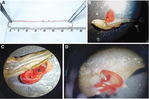
Parasite removed from a Danio rerio specimen measuring about 8 cm length. The cephalic extremity (right) presented a pinkish tone compared with the rest of the reddish body. (B) Alive parasite prolapsed from a D. rerio specimen. (C, D) Closer view with the aid of a stereomicroscope indicating the disruption site, with live parasite externalization. |

Photomicrographs of an Eustrongylides spp. larvae (A). Cephalic extremity displaying neural rings (nr), cuticle layer (cl), and part of a vascular system (vs); (B) inner papillae (ip, dotted arrows) and outer papillae (op, arrow); (C) medial part of the larva body and detail of transversal striations; (D) caudal extremity of a female larva presenting the vulva (v) and immature circular reproductive structure (*) and anal canal (ac); (E) anterior extremity presenting cephalic glands (cg) and detail of the esophagus lumen (oe); (F) esophagus–intestinal transition. |