
- Title
-
Development of Biomaterials Based on Biomimetic Trace Elements Co-Doped Hydroxyapatite: Physical, In Vitro Osteoblast-like Cell Growth and In Vivo Cytotoxicity in Zebrafish Studies
- Authors
- Tithito, T., Sillapaprayoon, S., Pimtong, W., Thongbunchoo, J., Charoenphandhu, N., Krishnamra, N., Lert-Itthiporn, A., Maneeprakorn, W., Pon-On, W.
- Source
- Full text @ Nanomaterials (Basel)

Scheme for multi-trace elements co-doped hydroxyapatite (THA) powders obtained by precipitation reactions. |

XRD patterns of sHA (a) and THA (b) powders. |

Magnetization dependence on magnetic field strength. |

Nitrogen adsorption–desorption isotherms of sHA and THA. |

SEM (TEM inset) images (a) and EDS analysis (b) of sHA particles; (c,d) represent SEM (TEM inset) and EDS analysis of THA particles, respectively. |

SEM image of THA surface before (a) and after (b) immersing the scaffolds in SBF for 7 days; (c) reveals the magnified view of the precipitation showing the accumulative structure of the mineral apatite particles and EDS analysis (d). |
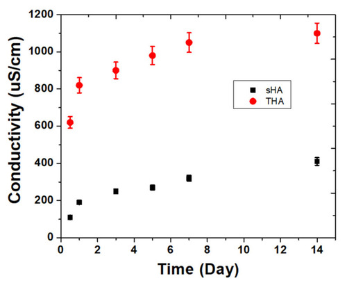
Electrical conductivity of the ions released from sHA and THA particles. |

SEM images of osteoblast cells on glass, sHA, and THA surfaces after 3, 5, and 7 days of culture. |
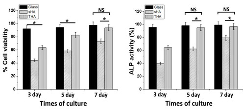
MTT assay and the relative alkaline phosphatase (ALP) activities of sHA and THA compared with the glass substrate (positive control; paired with each material) on days 3, 5, and 7. The control values were normalized to 100%. * p < 0.05 compared with the corresponding control group (mean ± SE). NS: no significance between the two groups. |

Survival rates for zebrafish embryos exposed to different concentrations (μg/mL) of sHA (a) and THA (b) at 72 h post-fertilization (hpf). Cumulative hatching rate of zebrafish embryos exposed to different concentrations of sHA (c) and THA (d) at 24, 48, and 72 hpf. The results are presented as mean ± SE (n = 3). a: p < 0.01 versus control. |

Digital photographs of zebrafish embryos at different stages of growth in solutions exposed to different concentrations (μg/mL) of sHA powders at 24, 48, and 72 hpf. |
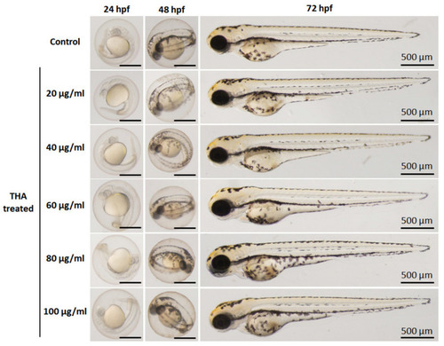
Digital photographs of zebrafish embryos at different stages of growth in solutions exposed to different concentrations (μg/mL) of THA powder at 24, 48, and 72 hpf. |

Effect of sHA and THA powders at varying concentrations on the heart rates of zebrafish embryos at 24 hpf (a) and 48 hpf (b). The results are presented as mean ± SE (n = 3), * p < 0.05 *** p < 0.001 versus control. |