
- Title
-
Grk7 but not Grk1 undergoes cAMP-dependent phosphorylation in zebrafish cone photoreceptors and mediates cone photoresponse recovery to elevated cAMP
- Authors
- Chrispell, J.D., Xiong, Y., Weiss, E.R.
- Source
- Full text @ J. Biol. Chem.
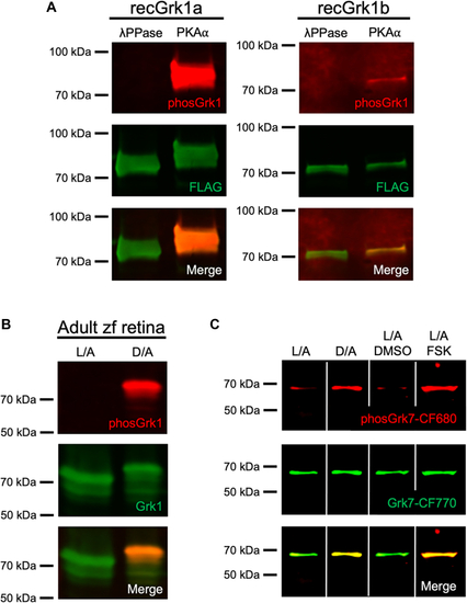
Immunospecificity of antibodies against phosphorylated Grk1 and Grk7.A, FLAG-tagged, purified recombinant zebrafish Grk1a and Grk1b treated in vitro with either λ phosphatase or PKA⍺ catalytic subunit were subjected to immunoblot analysis. Immunoblots were probed with antibodies against the FLAG-tag (green) and phosphorylated Grk1 (red) followed by incubation with secondary antibodies. B, Adult zebrafish were light- or dark-adapted, euthanized, and retinal homogenates subjected to immunoblot analysis. Immunoblots were probed with antibodies against Grk1 (green) and phosphorylated Grk1 (red), followed by incubation with secondary antibodies. C, Larvae at 5 dpf were light- or dark- adapted, followed by incubation with forskolin or vehicle (dimethyl sulfoxide [DMSO]) for 30 min. Larvae were euthanized and intact eyes were harvested for immunoblot analysis. Immunoblots were probed against Grk7 (green) and phosphorylated Grk7 (red) with anti-Grk7 antibody directly conjugated to CF770 and anti-phosphorylated Grk7 antibody directly conjugated to CF680. Panel C was spliced for clarity due to noncontiguous loading on the same gel. EXPRESSION / LABELING:
|
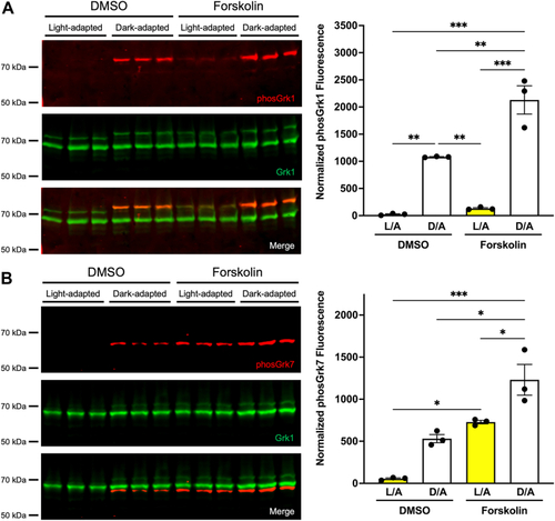
Effect of light exposure and forskolin incubation on phosphorylation of retinal Grks in WT zebrafish larvae at 5 dpf. Larvae were light- or dark-adapted, followed by incubation with forskolin or vehicle (DMSO, dimethyl sulfoxide) for 30 min. Larvae were euthanized and intact eyes were harvested for immunoblot analysis. Immunoblots were probed with antibodies against total Grk1 (green) and either (A) phosphorylated Grk1 (red) or (B) phosphorylated Grk7 (red) followed by incubation with secondary antibodies. The levels of phosphorylated Grk1 or Grk7 were normalized against total Grk1 and quantified. Statistical comparison of multiple groups was performed using a two-way ANOVA, followed by a Tukey post hoc test. Error bars represent SEM (n = 3). p ≤ 0.05 (∗), p ≤ 0.01 (∗∗), p ≤ 0.001 (∗∗∗). |
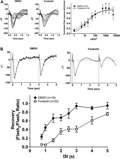
Electrophysiological light responses, normalized sensitivity, and recovery of the cone-mass receptor potential in forskolin-treated WT larvae at 5 dpf.A, Representative electroretinogram (ERG) traces of isolated cone-mass receptor potentials in larvae incubated in vehicle (DMSO, dimethyl sulfoxide) or forskolin for 25 min, followed by a 5-min coincubation with 2-amino-4-phosphonobutyric acid (APB). Reponses were recorded under dark-adapted conditions to 20-ms flashes of light of increasing intensities from 0.1 cd/m2 to 5000 cd/m2. The fast initial positive deflection is attributed to a photovoltaic effect with the recording microelectrode. Mean-normalized peak response amplitudes were fit using the Naka–Rushton function. B, Representative ERG waveforms of treated larvae subjected to successive stimuli using a dual flash paradigm of a 20-ms flash of saturating white light (1000 cd/m2) with an interstimulus interval (ISI) of 3 s. Vertical black dotted lines indicate time of stimulus. Cone-mass receptor potential recovery was plotted as the ratio of the maximum isolated cone mass receptor potential response of the second stimulus to that of the initial stimulus for ISIs ranging from 0.75 s to 5 s. A linear mixed model analysis of covariance found a significant effect of forskolin compared to vehicle [F(1, 350) = 115.6; p < 0.0001]. Error bars represent SEM. PHENOTYPE:
|

Recovery of the cone-mass receptor potential in grk KO zebrafish larvae treated with forskolin. Treated larvae were subjected to successive stimuli using a dual flash paradigm of a 20-ms flash of saturating white light (1000 cd/m2) with an interstimulus interval (ISI) ranging from 0.75 s to 5 s. Cone-mass receptor potential recovery was plotted as the ratio of the maximum isolated cone mass receptor potential response of the second stimulus to that of the initial stimulus for (A) grk1b−/− and (B) grk7a−/− larvae. A linear mixed model analysis of covariance found a significant effect of forskolin compared to vehicle (DMSO, dimethyl sulfoxide) for grk1b−/− (F(1, 224) = 17.82; p <0.0001) larvae, but not grk7a−/− larvae (F(1, 340) = 2.363; p = 0.1252). Error bars represent SEM. |

Grk1 phosphorylation in rod grk1a and cone grk1b KO zebrafish. Immunoblots were probed with antibodies against total Grk1 (green) and phosphorylated Grk1 (red), followed by incubation with secondary antibodies. Comparison of Grk1 phosphorylation in (A) light- or dark-adapted WT and grk1b−/− larvae at 5 dpf, (B) light- or dark-adapted WT and grk1b−/− adults, and (C) light- or dark-adapted WT, grk1b−/−, and grk1a−/− larvae at 5 dpf. Black arrowhead: Grk1a; Black arrow: Grk1b; White arrowhead, phosphorylatedGrk1a. Panel B was spliced for clarity due to noncontiguous loading on the same gel. |

Effect of light exposure and forskolin incubation on phosphorylation of retinal GRKs in grk1 KO zebrafish larvae at 5 dpf.A, grk1b−/− or (B) grk1a−/− larvae were light or dark adapted, followed by incubation with forskolin or vehicle (dimethyl sulfoxide [DMSO]) for 30 min. Larvae were euthanized and intact eyes were harvested for immunoblot analysis. Immunoblots were probed with antibodies against total Grk1 (green) and phosphorylated Grk1 (red) followed by incubation with secondary antibodies. The levels of phosphorylated Grk1 were normalized against total Grk1 and quantified. Statistical comparison of multiple groups was performed using a two-way ANOVA followed by a Tukey post hoc test. Error bars represent SEM (n = 3). p ≤ 0.05 (∗), p ≤ 0.01 (∗∗), p ≤ 0.001 (∗∗∗), p ≤ 0.0001 (∗∗∗∗). |
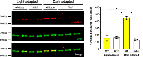
Grk1 phosphorylation in Nrl−/− mice. Adult mice were light or dark adapted, euthanized, and retinas harvested. Immunoblots were probed with antibodies against total Grk1 (green) and phosphorylated Grk1 (red), followed by incubation with secondary antibodies. The levels of phosphorylated Grk1 were normalized against total Grk1 and quantified. Statistical comparison of multiple groups was performed using a two-way ANOVA followed by a Tukey post hoc test. Error bars represent SEM, (n=2–3). p ≤ 0.05 (∗). |
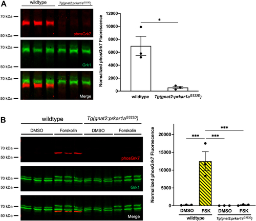
Dark-adapted Grk phosphorylation in 5 dpf transgenic Tg(gnat2:prkar1aG323D) zebrafish larvae expressing PKA dominant negative RI⍺B in cones. Larvae were (A) dark adapted for 15 min or (B) light adapted, followed by incubation with forskolin or vehicle (DMSO, dimethyl sulfoxide) for 30 min, then euthanized and intact eyes were harvested for immunoblot analysis. Immunoblots were probed with antibodies against total Grk1 (green) and phosphorylated Grk7 (red), followed by incubation with secondary antibodies. The levels of detectable phosphorylated Grk7 were normalized against total Grk1 and quantified. Statistical comparison of multiple groups was performed using a Student's t test. Error bars represent SEM (n = 3). p ≤ 0.05 (∗). |