
- Title
-
Aluminum (Al) causes a delayed suppression of nucleotide excision repair (NER) capacity in zebrafish (Danio rerio) embryos via disturbance of DNA lesion detection
- Authors
- Paul, G.V., Huang, Y.Y., Wu, Y.N., Ho, T.N., Hsiao, H.I., Hsu, T.
- Source
- Full text @ Ecotoxicol. Environ. Saf.

Fig. 1. Effects of increasing Al concentration on the survival of developing zebrafish. Zebrafish embryos at 1 hpf were exposed to Al at the indicated concentrations until the larval stage at 96 hpf. Exposure was carried out by individually transferring embryos to the wells of 24-well plates with 2 mL of test solution per well and endpoints described in OECD guideline TG 236 were used as the indicators for lethal responses. Each bar graph represents the percentage survival rate of embryos or larvae as the mean and standard deviation of four separate experiments. * indicates p < 0.05 and ** indicates p < 0.01 when compared to the survival rate of nonexposed embryos at the same developmental stage. |
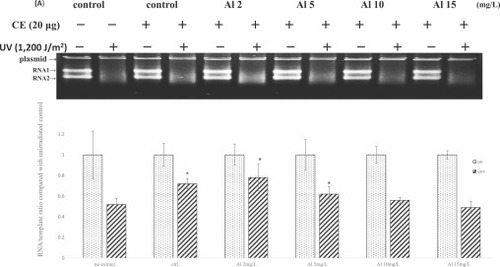
Fig. 2. Downregulation of NER capacity in zebrafish embryos exposed to sublethal levels of Al. Embryos at 1 hpf were exposed to Al at the indicated concentrations for 71 h and NER capacity was detected by a transcription-based repair assay. (A) A representative gel showing the results of in vitro transcription promoted by 20 μg zebrafish extract proteins at 37 °C for 3 hr. (B) Quantitative determination of the effects of Al on NER capacity in zebrafish embryos. Each bar graph represents the mean and standard deviation of five separate experiments. *indicates p < 0.05 when compared to protein-free negative control. CE stands for crude extracts. No extract under the first two bar graphs indicates the transcriptional activities of marker cDNA in UV-irradiated and unirradiated plasmid not incubated with extract proteins. |
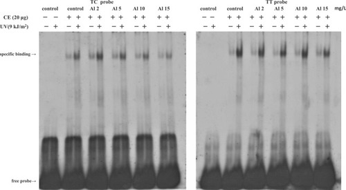
Fig. 3. Differential effects of Al on CPD and 6–4PP binding activities in zebrafish embryos. Embryos at 1 hpf were treated with Al at the indicated concentrations for 71 h. UV(9 kJ/m2)-irradiated or unirradiated TT or TC probe (6 pmole) was incubated at 30 °C for 30 min with zebrafish extracts containing 20 μg proteins and the formation of damage-dependent binding complexes was detected by EMSA. CE stands for crude extracts. |
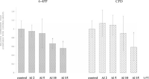
Fig. 4. Quantitative analysis of the effects of Al on CPD and 6–4PP sensing activities in zebrafish embryos. Each bar graph represents the mean and standard deviation of 4–6 independent experiments. * indicates p < 0.05 when compared to control. |

Fig. 5. Delayed response of NER capacity in zebrafish embryos to Al. Embryos at 1 hpf were treated with Al at 15 mg/L for the indicated time periods and UV damage repair activities in the extracts of stressed and unstressed embryos at the same developmental stages were determined. (A) A representative gel showing the results of transcription-based repair assay. (B) Time-course effects of Al on NER capacity determined by quantitative analysis. Each bar graph represents the mean and standard deviation of five independent experiments. * indicates p < 0.05 when compared to protein-free negative control. CE stands for crude extracts. |

Fig. 6. Exposure time-dependent effects of Al on DNA damage recognition activities in zebrafish embryos. Embryos at 1 hpf were exposed to Al at 15 mg/L for different time periods and 6–4PP detection activities in Al-treated and untreated embryos at the same developmental stages were examined by EMSA. (A) A representative gel image showing the effects of Al on 6–4PP sensing activities after different time periods of exposure. (B) Quantitative determination of 6–4PP binding activities in embryos exposed to Al for different time periods. (C) Time-course effects of Al on ddb2 and xpc expression analyzed by real-time RT-PCR. Each bar graph represents the mean and standard deviation of 3–4 separate experiments. *, ** and *** indicate p < 0.05, p < 0.01and p < 0.005, respectively. CE stands for crude extracts. |

Fig. 7. Inhibition of lesion incision/excision in UV-irradiated embryos preexposed to Al. Embryos preexposed to Al at 15 mg/L for 35 h and nonexposed embryos were separately irradiated with UV (1200 J/m2) to induce NER. The levels of excised oligonucleotides were detected by TdT-mediated end labeling assay. Arrows indicate the positions of 26- and 32-mer size markers. (A) A representative gel image showing the levels of excised oligomers in embryos under each experimental condition. (B) Quantitative analysis of the effects of Al on lesion incision/excision. Each bar graph represents the mean and standard deviation of three separate experiments. The levels of short oligonucleotides in UV-irradiated embryos preexposed to Al were significantly (p < 0.01) lower than those in UV-irradiated embryos nonexposed to Al. |