
- Title
-
Expression of Nerve Growth Factor and Its Receptor TrkA in the Reproductive System of Adult Zebrafish
- Authors
- Cacialli, P.
- Source
- Full text @ Vet Sci
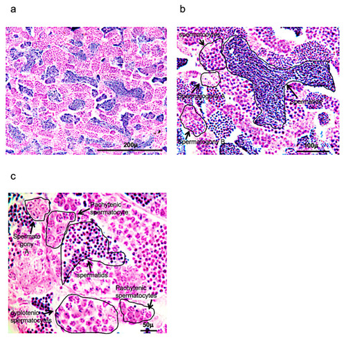
Hematoxylin-eosin staining of adult zebrafish testis. (a) Overview of adult zebrafish testis by hematoxylin-eosin staining. (b) Spermatogony A-B, spermatocytes and spermatids in adult zebrafish testis by hematoxylin-eosin staining. (c) High magnification of spermatogony A, spermatocytes (different phases of meiosis) and spermatids in adult zebrafish testis by hematoxylin-eosin staining. Scale bars are: 200 µ (a); 100 µ (b); and 50 µ (c). |
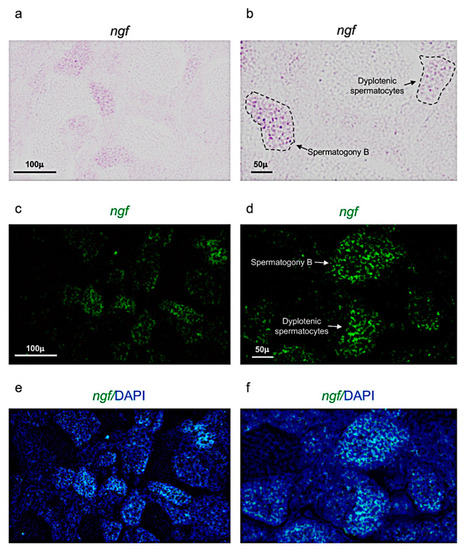
Ngf expression pattern in adult zebrafish testis. (a,b) Chromogenic in situ hybridization of ngf in adult zebrafish testis. (c,d) Fluorescence in situ hybridization of ngf in adult zebrafish testis. (e,f) Fluorescence in situ hybridization of ngf and cell nuclei (DAPI), in adult zebrafish testis. Scale bars are: 100 µ (a,c,e); 50 µ (b,d,f). EXPRESSION / LABELING:
|
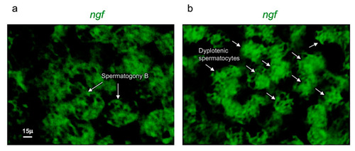
Ngf is specifically expressed in spermatogony B and dyplotenic spermatocytes. High magnification, fluorescence in situ hybridization of ngf, specifically expressed in (a) spermatogony B and in (b) dyplotenic spermatocytes in adult zebrafish testis. Scale bar: 15 µ (a,b). EXPRESSION / LABELING:
|
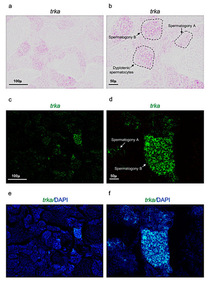
Trka expression pattern in adult zebrafish testis. (a,b) Chromogenic in situ hybridization of trka in adult zebrafish testis. (c,d) Fluorescence in situ hybridization of trka in adult zebrafish testis. (e,f) Fluorescence in situ hybridization of trka and cell nuclei (DAPI) in adult zebrafish testis. Scale bars: 100 µ (a,c,e); 50 µ (b,d,f). EXPRESSION / LABELING:
|
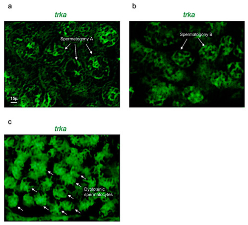
Trka is specifically expressed in spermatogony A, B and dyplotenic spermatocytes. High magnification, fluorescence in situ hybridization of ngf, specifically expressed in (a) spermatogony A; (b) spermatogony B; and (c) dyplotenic spermatocytes in adult zebrafish testis. Scale bar: 15 µ (a–c). EXPRESSION / LABELING:
|

Hematoxylin-eosin staining of adult zebrafish ovary. (a) Overview of adult zebrafish ovary by hematoxylin-eosin staining, five oocyte stages (I-II-II-IV-V). (b) Oocyte stages II and V. (c) Oogonia characterized by large euchromatic germinal vescicle (GV), and several nucleoli peripherically located. (d) Oocyte during primary growth (stages II–III), characterized by an increase of nucleoli in GV, and in ooplasma were present oil droplets (od) around the GV. (e) Oocyte (stages III) characterized by numerous nucleoli at the periphery of GV. (f) numerous oil droplets and cortical alveoli (ca). (g) The oocyte (stages IV) is enveloped by zona pellucida (zp) and a single layer of follicular cells (fc). (h) Oocyte (stage V) characterized by a significant increase of number and size of the yolk globules (yg). Significant increase in the thickness of the zona pellucida and appearance of thecal layer. Scale bars are: 100 µ (a,b); 60 µ (d–f); 20 µ (c,g,h). |
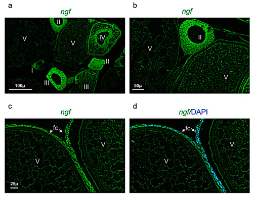
Ngf expression pattern in adult zebrafish ovary. (a) Fluorescence in situ hybridization of ngf in adult zebrafish ovary, mainly expressed in oocyte at different stages: II-III-IV and in follicular cells of stage V. (b) Fluorescence in situ hybridization of ngf in oocytes at II and V stages. (c) Fluorescence in situ hybridization of ngf, high magnification of follicular cells in oocyte at stage V. (d) Fluorescence in situ hybridization of ngf and cell nuclei (DAPI), high magnification of follicular cells in oocyte at stage V. Scale bars: 100 µ (a); 50 µ (b); and 25 µ (c,d). EXPRESSION / LABELING:
|
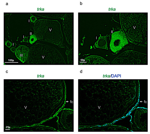
Trka expression pattern in adult zebrafish ovary. (a) Fluorescence in situ hybridization of trka in adult zebrafish ovary, mainly expressed in oocytes at different stages: II, III, and in follicular cells of stage V. (b) Fluorescence in situ hybridization of trka in oocytes at the stages I, II, and V. (c) Fluorescence in situ hybridization of trka, high magnification of follicular cells in oocyte at stage V. (d) Fluorescence in situ hybridization of ngf and cell nuclei (DAPI), high magnification of follicular cells in oocyte at stage V. Scale bars: 100 µ (a); 50 µ (b); and 25 µ (c,d). EXPRESSION / LABELING:
|