FIGURE SUMMARY
Title

Excess DHA Induces Liver Injury via Lipid Peroxidation and Gut Microbiota-Derived Lipopolysaccharide in Zebrafish

Authors
Ding, Q., Hao, Q., Zhang, Q., Yang, Y., Olsen, R.E., Ringø, E., Ran, C., Zhang, Z., Zhou, Z.
Source
Full text @ Front Nutr

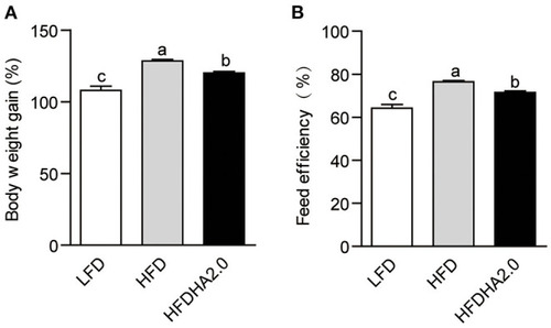
The effects of DHA supplementation on growth and feed efficiency in zebrafish. (A) Body weight gain and (B) feed efficiency of LFD, HFD, or HFDHA2.0-fed zebrafish. Values are means ± SEMs (n = three biological replicates). The mean values without a common letter are significantly different, p < 0.05. LFD, low-fat diet; HFD, high-fat diet; HFDHA2.0, 2% DHA-supplemented HFD.

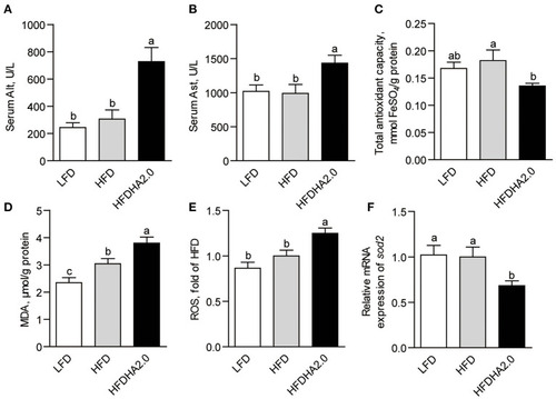
The effects of DHA on liver injury and oxidative stress in zebrafish. The activities of serum (A) Alt and (B) Ast of LFD, HFD or HFDHA2.0-fed zebrafish. Hepatic (C) T-AOC, (D) MDA and (E) ROS of LFD, HFD or HFDHA2.0-fed zebrafish. The relative mRNA expression of (F)sod2 of LFD, HFD, or HFDHA2.0-fed zebrafish. The values are means ± SEMs (n = 5 or 6 biological replicates). The mean values without a common letter are significantly different, p < 0.05. LFD, low-fat diet; HFD, high-fat diet; HFDHA2.0, 2% DHA-supplemented HFD.

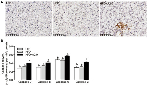
The effects of DHA on liver apoptosis in zebrafish. (A) Tunnel staining of liver sections of LFD, HFD, or HFDHA2.0-fed zebrafish. The scale bar is 50 μm. (B) The activities of caspase-9, caspase-8, caspase-6, and caspase-3 in LFD, HFD, or HFDHA2.0-fed zebrafish. The values are means ± SEMs (n = 6 biological replicates). The mean values without a common letter are significantly different, p < 0.05. LFD, low-fat diet; HFD, high-fat diet; HFDHA2.0, 2% DHA-supplemented HFD.

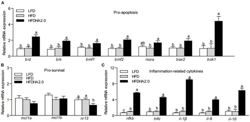
The effects of DHA on the relative mRNA expression of Bcl-2 family and inflammation-related cytokines in the livers of zebrafish. (A) The relative mRNA expression of pro-apoptotic proteins of LFD, HFD, or HFDHA2.0-fed zebrafish. (B) The relative mRNA expression of pro-survival proteins of LFD, HFD, or HFDHA2.0-fed zebrafish. (C) The relative mRNA expression of inflammatory cytokines of LFD, HFD, or HFDHA2.0-fed zebrafish. The values are means ± SEMs (n = 4–6 biological replicates). The mean values without a common letter are significantly different, p < 0.05. LFD, low-fat diet; HFD, high-fat diet; HFDHA2.0, 2% DHA-supplemented HFD.

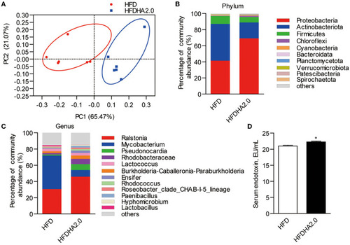
The effects of DHA supplementation on gut microbial community in zebrafish. (A) PCoA analysis of gut microbiota in HFD- or HFDHA2.0-fed zebrafish. (B) Relative abundance at the phylum level of gut microbial community from HFD- or HFDHA2.0-fed zebrafish. (C) Relative abundance at the genus level of gut microbial community from HFD- or HFDHA2.0-fed zebrafish. (D) Serum endotoxin in HFD- or HFDHA2.0-fed zebrafish. The values are means ± SEMs (n = 4 or 6 biological replicates). *p < 0.05. HFD, high-fat diet; HFDHA2.0, 2% DHA-supplemented HFD.

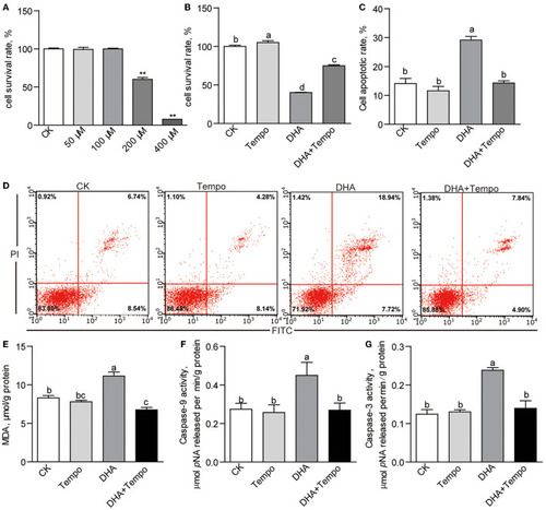
The effects of antioxidant, 4-hydroxy-Tempo (Tempo), on DHA cytotoxicity in ZFL cells. (A) The cell survival rates of ZFL cells treated with increasing concentrations of DHA for 24 h. (B) The cell survival rates and (C) the cell apoptotic rates of ZFL cells co-treated with DHA and tempo for 24 h. (D) The representative images of cell apoptosis of ZFL cells co-treated with DHA and tempo for 24 h. (E) The MDA levels in ZFL cells co-treated with DHA and tempo for 24 h. The activities of (F) caspase-9 and (G) caspase-3 in ZFL cells co-treated with DHA and tempo for 24 h. Values are means ± SEMs (n = 3 ~ 8 biological replicates). The mean values without a common letter are significantly different, p < 0.05. **p < 0.01.

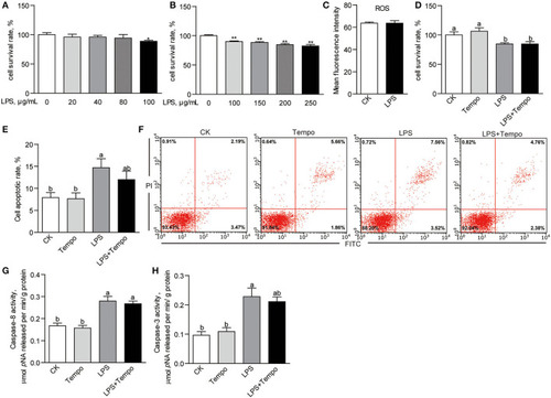
The effects of LPS on cell viability and apoptosis of ZFL cells. (A) The cell survival rates of ZFL cells treated with lower concentrations of LPS for 24 h. (B) The cell survival rates of ZFL cell treated with higher concentrations of LPS for 24 h. (C) The intracellular ROS of ZFL cells treated with 100 μg/ml LPS for 24 h. (D) The cell survival rates and (E) cell apoptotic rates of ZFL cells co-treated with LPS and tempo for 24 h. (F) The representative images of cell apoptosis of ZFL cells co-treated with LPS and tempo for 24 h. The activities of (G) caspase-8 and (H) caspase-3 in ZFL cells co-treated with DHA and tempo for 24 h. The values are means ± SEMs (n = 3–8 biological replicates). The mean values without a common letter are significantly different, p < 0.05. *p < 0.05, **p < 0.01.

Acknowledgments
This image is the copyrighted work of the attributed author or publisher, and ZFIN has permission only to display this image to its users. Additional permissions should be obtained from the applicable author or publisher of the image. Full text @ Front Nutr