FIGURE SUMMARY
Title

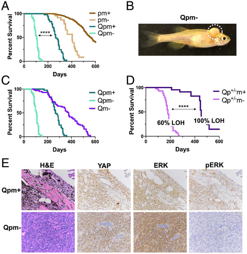
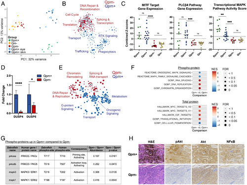
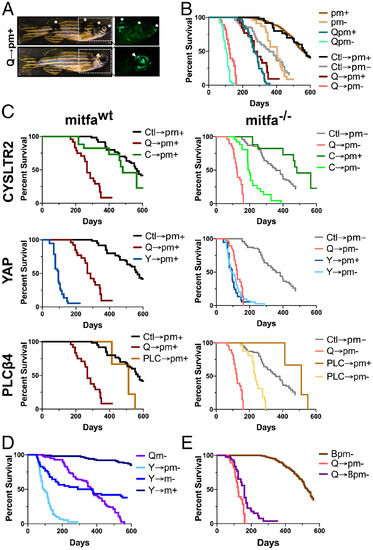
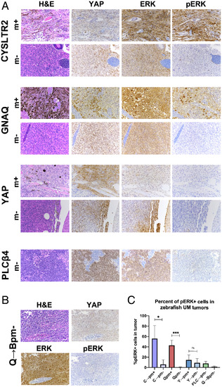
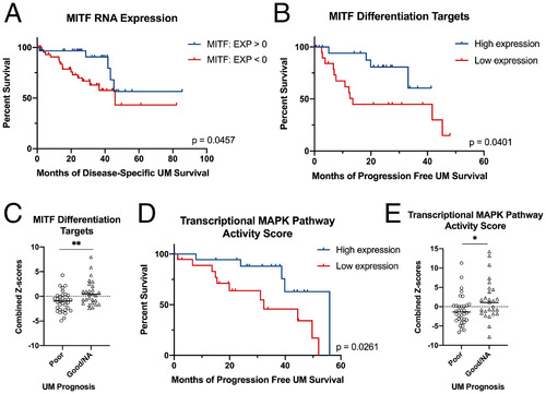
MITF deficiency accelerates GNAQ-driven uveal melanoma

Authors
Phelps, G.B., Hagen, H.R., Amsterdam, A., Lees, J.A.
Source
Full text @ Proc. Natl. Acad. Sci. USA

Mitfa loss accelerates GNAQQ209L-driven tumorigenesis, and resulting tumors stain negatively for hyperactive ERK; Q, Tg(mitfa:GNAQQ209L); p, tp53M214K/M214K; m+, mitfa+/+; m−, mitfa−/−; p+/−, tp53+/M214K. (A) Kaplan–Meier curves for the indicated genotypes show that overall survival of Tg(mitfa:GNAQQ209L)-expressing zebrafish was significantly decreased by mitfa deficiency (P < 0.0001, determined by log-rank test). The pm+ and pm− zebrafish develop MPNSTs, while Qpm+ and Qpm− zebrafish develop UMs and, less frequently, MPNSTs. (B) A representative image, with tumor outlined by dotted line, shows the typical unpigmented UM tumor phenotype of Qpm− zebrafish. (C) Kaplan–Meier curves show that mitfa−/− also cooperates with GNAQQ209L to decrease overall survival in a tp53-WT background. The Qpm+ and Qpm− curves from A are included for comparison. (D) Kaplan–Meier curves showing that Qp+/−m− zebrafish have significantly reduced survival compared to Qp+/−m+ counterparts (P < 0.0001, determined by log-rank test) as well as a reduced reliance on tp53 LOH, as determined by DNA sequencing of the mutation-bearing tp53 exon of excised tumors. (E) Representative images (n ≥ 3 for each stain and genotype) of hematoxylin and eosin (H&E) and IHC for YAP, ERK1/2, and phospho-ERK1/2 (pERK; active) for Qpm+ and Qpm− tumors. YAP activation was detected through nuclear localization.

RNA sequencing and phospho-proteomics determine that Qpm− tumors are deficient in PLCβ4–PKCδ/ε signaling and up-regulate MYC target genes. Genotype abbreviations are as indicated in Fig. 1; B, Tg(mitfa:BRAFV600E). (A–C) RNA-sequencing data were generated for the following tumor numbers, genotypes, and tumor locations: 9 Bpm+, 8 Qpm+ eye, 9 Qpm+ skin, 10 Qpm−, and 10 Qm− (GSE190802). (A) PCA shows distinct clustering of Qpm+ tumors (regardless of their anatomical location), Qpm− and Qm− (regardless of tp53 status), and Bpm+. (B) Cytoscape enrichment map shows GSEA c2cp_Reactome data sets that were significantly enriched (false discovery rate [FDR] q value < 0.05) in Qpm− (red) versus Qpm+ (blue). Each circle denotes a gene set, circle size denotes the number of genes in each gene set, and clustering and line length is determined by similar genes within each gene set. Complete GSEA results are shown in Dataset S1. (C) For each individual Bpm+, Qpm+, Qpm−, and Qm− tumor, combined Z scores were calculated for the identified zebrafish orthologs for the MITF target gene (Left), PLCβ4 pathway (Middle), or MAPK transcriptional activity (Right) gene sets. The bar denotes the medians for each tumor type. Qpm− and Qm− tumors have significantly reduced expression compared with either Qpm+ or Bpm+ tumors in each of the three gene sets (P < 0.001, determined by Student’s unpaired t test). (D) qRT-PCR for the top two differentially expressed genes in the MAPK transcriptional activity gene set, DUSP4 and DUSP6, on new Qpm+ and Qpm− tumor samples (n = 6 each). Results are normalized to ACTB2 expression. Fold change is relative to the average of the Qpm+ samples, with error bars indicating SD. P < 0.0001 for DUSP4 and P = 0.033 for DUSP6, as determined by Student’s unpaired t test. (E–G) Phospho-peptides and total proteins from Qpm− and Qpm+ tumors (n = 5/genotype) were quantified by mass spectrometry, and GSEA was conducted (complete results are shown in Dataset S1. (E) Cytoscape enrichment map shows c2cp Reactome and c5 Biological Processes data sets significantly enriched (FDR q value < 0.05) in phospho-proteins from Qpm− (red) versus Qpm+ (blue) tumors. (F) Key gene sets identified as significantly different between Qpm− and Qpm+ for phospho-proteins (Top) or total protein (Bottom), demonstrating enrichment of MAPK signaling in Qpm+ and MYC and E2F targets in Qpm−; NES, normalized enrichment score. (G) Phospho-peptides associated with PKCδ, PKCε, ERK1, and ERK2 activation that were reduced in Qpm− compared to Qpm+. (H) Representative IHC images (from n ≥ 3 per stain and genotype) for phospho-Akt (p-AKT; active), total AKT, and NF-κB (with nuclear staining indicating the active form) show that Qpm− tumors are deficient for signaling from pathways other than MAPK downstream of PKC–RasGRP3.

EXPRESSION / LABELING:
Antibodies:
Fish:
Anatomical Term:
Stage: Adult

A mosaic expression system determines that CYSLTR2L129Q, YAPAA, and PLCβ4D630Y can drive UM tumorigenesis. Mosaic zebrafish are denoted by X➛Y, where X is the gene introduced, and Y is the recipient genotype; Ctl, control GOI–GFP vector; Q, GNAQQ209L; C, CYSLTR2 L129Q; Q, GNAQQ209L; Y, YAPAA; PLC, PLCB4D630Y. Germline zebrafish lines and recipient genotypes are as indicated in Figs. 1 and 2. (A) Representative zebrafish images confirm that introduction of GNAQQ209L via the GOI–GFP vector results in GFP+ pigment patches and tumors. Asterisks and arrows denote pigment patches corresponding to GFP expression. (B) Kaplan–Meier curves show that mosaic zebrafish with control vector or GNAQQ209L introduced into pm+ or pm− recipients all have overall survivals that closely mirror their germline pm+, pm−, Qpm+, and Qpm− equivalents (P = n.s.). As with the germline mutants, Q➛pm+ versus Ctl➛pm+ and Q➛pm− versus Ctl➛pm− were both significantly different (P < 0.001, as determined by log-rank test). (C) Kaplan–Meier curves show the effects of mosaic expression of CYSLTR2L129Q, YAPAA, and PLCβ4D630Y in tp53-mutant zebrafish that are either mitfa+/+ (Left) or mitfa−/− (Right). Q➛pm+, Q➛pm−, Ctl➛pm+, and Ctl➛pm− data from B are shown for comparison. Statistical significance is as follows: CYSLTR2, C➛pm+ versus Ctl➛pm+ n.s., C➛pm− versus Ctl➛pm− P < 0.0001, as determined by log-rank test; YAP, Y➛pm+ versus Ctl➛pm+ P < 0.0001, Y➛pm− versus Ctl➛pm− P < 0.0001, as determined by log-rank test; PLCβ4, PLC➛pm+ versus Ctl➛pm+ n.s., PLC➛pm− versus Ctl➛pm− P < 0.0001, as determined by log-rank test. (D) Kaplan–Meier curves show that YAP cooperates with mitfa−/− in a tp53-WT background. Overall survival of Y➛m+ versus YAP➛m−, significant to P < 0.0001, as determined by log-rank test. (E) Kaplan–Meier curves of Q➛Bpm− versus Bpm− controls, with Q➛pm− from B shown for comparison, show that GNAQQ209L is dominant over BRAFV600E in allowing tumors to form the mitfa−/− background. Statistical significance of overall survival is P < 0.0001 for Q➛Bpm− versus Bpm− and P = 0.0003 for Q➛Bpm− versus Q➛pm−, as determined by log-rank test.

CYSLTR2L129Q-, YAPAA-, and PLCβ4D630Y-driven tumors do not display hyperactive ERK staining in the mitfa−/− context. Genotype abbreviations are as described in Fig. 3. (A and B) Representative images of H&E staining and IHC for YAP, ERK, or phospho-ERK (pERK) on the indicated tumor genotypes. YAP activation is determined by nuclear localization and ERK activation by phospho-ERK. (A) C➛pm+ tumors and Qpm+ tumors are positive for nuclear YAP and phospho-ERK, while C➛pm−, Qpm−, and PLC➛pm− tumors display only YAP activation; n ≥ 3 for all genotypes and stains, except n = 2 for each Y➛pm− and C➛pm+ IHC stains. (B) Q➛Bpm− displays nuclear YAP but not phospho-ERK; n = 5 for all stains. (C) Graph shows the percent of UM tumor cells positive for phospho-ERK quantified by QuPath (n ≥ 3 for all genotypes, except n = 2 for Y➛pm− and C➛pm+), and error bars denote SD. Statistical significance is as follows: C➛pm+ versus C➛pm− P = 0.0435, Q➛pm+ versus Q➛pm− P = 0.0001, Y➛pm+ versus Y➛pm− P = 0.551, as determined by Student’s unpaired t test.

Decreased expression of MITF target genes and decreased MAPK transcriptional activity correlate with poor UM patient survival. RNA sequencing of primary human UM (n = 80 patients) and the corresponding survival data were obtained from TCGA PanCancer Atlas database. (A) Patients with relatively lower MITF RNA expression (z score < 0; n = 49) have significantly decreased (P = 0.0457, determined by log-rank test) disease-free survival compared to patients with relatively higher MITF RNA expression (z score > 0; n = 31). (B) Z scores for the MITF differentiation target genes DCT, TYR, TYRP1, and SILV were calculated and summed for each UM tumor, and patients in the lower quartile (n = 20) were found to have significantly decreased (P = 0.04, determined by log-rank test) progression-free survival compared to patients in the upper quartile (n = 20). (C) UM patients were binned by “poor prognosis” (those deceased or with progression of the disease [n = 34]) versus “good/NA prognosis” (those last reported as living without progression for at least 2 y [n = 27]). The poor prognosis cohort showed significantly lower combined z scores for the four MITF differentiation targets (P = 0.0041, determined by Student’s unpaired t test; line denotes median). (D) Z scores for genes in the transcriptional MAPK activity gene set were calculated and summed. Patients in the lower quartile (n = 20) showed significantly decreased (P = 0.0261, determined by log-rank test) progression-free survival compared to patients in the upper quartile (n = 20). (E) UM patients with poor prognosis (binned as described in C) have a significantly lower transcriptional MAPK pathway activity combined z scores (P = 0.0218, determined by Student’s unpaired t test; line denotes median) than the good/NA prognosis cohort.

Acknowledgments
This image is the copyrighted work of the attributed author or publisher, and ZFIN has permission only to display this image to its users. Additional permissions should be obtained from the applicable author or publisher of the image. Full text @ Proc. Natl. Acad. Sci. USA