
- Title
-
Colorimetric and fluorescent TRAP assays for visualising and quantifying fish osteoclast activity
- Authors
- Ethiraj, L.P., Fong, E.L.S., Liu, R., Chan, M., Winkler, C., Carney, T.J.
- Source
- Full text @ Eur. J. Histochem.

Identification of activated osteoclasts using conventional TRAP stain. A-H) Brightfield images of zebrafish (A-D) and medaka (E-H) adult fins (A-B,E,F), or 12dpf larvae (C,D,G H) processed by colorimetric TRAP staining. Individuals are either wild-type (WT; A, C, E, F), homozygous csf1raj4e1 mutants (B,D) or transgenic for rankl:HS:cfp (G,H). Images are of whole-mount fins at 4 dpc (A,B), whole-mount ventral views (C, D) or longitudinal (E) or transverse cryosections (G,H). Fins in (E,F) have been amputated and stained immediately (0dpa; E) or after 6 days (dpa; F). The larva in (H) has been subjected to heat shock to express RANKL, while the larva in (G) was not heat shocked. Red precipitate (blue arrowheads) indicates TRAP locations at fractures (A), pharyngeal teeth of the fifth ceratobranchial bone (C), in regenerating fin rays at 6 days post amputation (6dpa; F) or around notochord in larvae with excess RANKL induced by heat shock (H). TRAP staining is absent when there no RANKL overexpression (G), at the start of fin regeneration (0dpa; E) or following genetic ablation of osteoclasts (B, D; open blue arrowheads), indicating specificity. Scale bars: A) 200 μm; C) 100 μm; F,H) 50 μm. |

A fluorescent TRAP method labels osteoclast activity in zebrafish bone fractures. A-L) Whole-mount widefield fluorescent images of adult fin ray fractures processed for ELF97 fluorescence. Fins were fixed at 3 dpc (A-C), 7 dpc (D-F), 14 dpc (G-I) and 21 dpc (J-L), and were either WT (A,D,G,J), homozygous csf1raj4e1 mutants (B, E, H, K) or treated with 100 μg/mL bisphosphonate (+BP; C, F, I, L). M: Mean fluorescence of crush sites at given timepoints and under the three conditions. ***= p<0.001, *=p<0.05, (ANOVA with Bonferroni multiple comparisons test, n=12). N: Mean ELF97 fluorescence at the crush sites of 1 dpc WT zebrafish following storage for 0, 1, 5, 7, 14, 21 days. There was no statistically significant variation over the course of 21 days of storage compared against initial staining (ANOVA, Tukey's multiple comparisons test, n=12). Scale bar: A) 100 μm. |
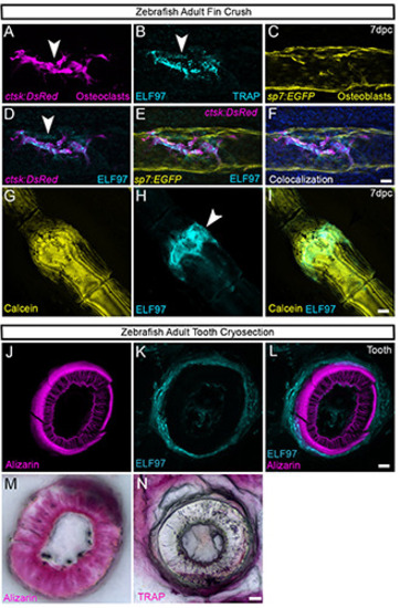
Colocalization of TRAP-ELF97 reaction product with fluorescent proteins and dyes. A-F) Whole-mount confocal images of a fin ray fracture in ctsk:Dsred; sp7:egfp double transgenic adults, processed at 7 dpc for ELF97 TRAP fluorescence and counterstained with DAPI. Individual fluorescent channels for ctsk:Dsred (A), ELF97 (B), sp7:egfp (C), DsRed and ELF97 channel overlay (D), DsRed, EGFP and ELF97 channel overlay (E), all channels including DAPI (F). TRAP-ELF97 fluorescence is seen associated with DsRed labelled osteoclasts within the callus. G-I) Confocal images of whole-mount adult zebrafish fin ray subjected to crush fracture and processed at 4 dpc for Calcein (G) and ELF97 (H) staining. TRAP activity is seen within the bone matrix of the callus (I). J-N) Confocal image of cryosection through an adult zebrafish pharyngeal tooth which has been stained with Alizarin Red (J,M), ELF97 (K) and colorimetric TRAP (N). Overlay of both channels indicates TRAP staining surrounding the bone mineral (L). Scale bars: F,I,L,N) 20 μm. |

Fluorescent TRAP staining can be combined with immunofluorescence. A-L) Confocal images of dissected fifth ceratobranchial bone with pharyngeal teeth of a ctsk:DsRed; sp7:EGFP double transgenic adult which had been processed for ELF97 TRAP staining (C,H) and immunostained with antibodies against EGFP (A,G) and DsRed (B,I). Both whole-mount (A-F) and cryosectioned (G-L) samples are presented. Co-localisation demonstrates ELF97 signal associated with DsRed positive osteoclasts at the base of the teeth (D,E,J,K), whereas eGFP positive osteoblasts are found within the teeth (E, F, K, L). Fluorescent channels are also shown with DIC image (F,L). Scale bars: F) 200 μm; L) 50 μm. |