
- Title
-
Modeling Human Cardiac Arrhythmias: Insights from Zebrafish
- Authors
- Gauvrit, S., Bossaer, J., Lee, J., Collins, M.M.
- Source
- Full text @ J Cardiovasc Dev Dis
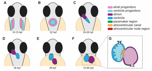
Stages of heart development in zebrafish from 10 to 96 h post-fertilization (hpf). (A) Atrial and ventricular cardiac progenitors are located in the anterior lateral plate mesoderm by ~15 hpf. (B) The cardiac disc is visible by 22 hpf as cardiac progenitors surround endocardial cells at the midline. (C) At 24 hpf, the linear heart tube forms and jogs to the left in preparation for heart looping. (D) At 36 hpf, the heart tube undergoes rightward looping. The AV canal (orange) begins to develop between the cardiac chambers. (E) At 48 hpf, the cardiac chambers begin to balloon and expand outwards. The bulbus arteriosus (dark blue) and AV canal (orange) continue to develop and mature. (F) From 72 to 96 hpf, the cardiac chambers expand and align beside each other. (G) A cross-section of a 96 hpf heart showing the trabeculae, the finger-like muscular projections on the inner wall of the ventricle, and endocardial leaflets (orange) of the AV canal. (A–D) dorsal views; (E–G) ventral views. |
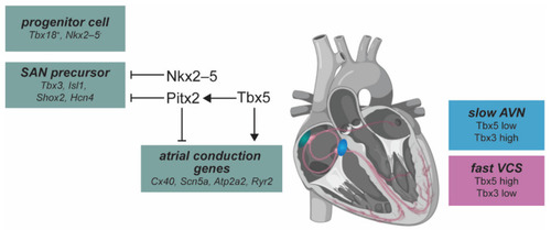
Overview of molecular pathways in the mammalian cardiac conduction system. The sinoatrial node (SAN, green) develops from Tbx18+, Nkx2-5low progenitor cells. Nkx2-5 represses expression of Tbx3 and Isl1 to establish the boundary between the SAN and atrial cardiomyocytes. SAN precursor cell differentiation is marked by expression of Tbx3, Isl1, Shox2, and Hcn4. Pitx2 represses SAN development on the left side of the sinus venosus by repressing this transcriptional network. The Tbx5 (activator) and Pitx2 (repressor) regulatory loop regulates atrial conduction genes. Relative Tbx3/Tbx5 dosage determines specification of the conduction system. The atrioventricular node (AVN, blue) acts as a secondary pacemaker, and is characterized by slow conduction that is patterned by low levels of Tbx5 and high levels of Tbx3. A fast conduction state in the ventricular conduction system (VCS, pink) is specified by higher levels of Tbx5 and low Tbx3. |