
- Title
-
Two-photon scanned light sheet fluorescence microscopy with axicon imaging for fast volumetric imaging
- Authors
- Lin, P.Y., Hwang, S.L., Lee, C.H., Chen, B.C.
- Source
- Full text @ J. Biomed. Opt.

(a) Schematic of 2p LSFM with axicon imaging. Inset shows the extended DOF using axicon imaging. Measured PSFs (b) without and (c) with axicon imaging. (d) The corresponding intensity line profiles of measured PSF and lateral FWHM factor at different axial positions: lateral (left), axial (upper right); lateral FWHM factor (lower right). Red: with axicon imaging. Blue: without axicon imaging. HWP, half-wave plate; PBS, polarization beam splitter; L1 to L9, achromatic doublet lens; and TL, tube lens. Scale bars: |

Imaging fluorescent beads by 2p LSFM (a) with and (b) without axicon imaging. (c) Normalized intensity line profile through a fluorescent bead. Red: with axicon imaging. Blue: without axicon imaging. Scale bars: |
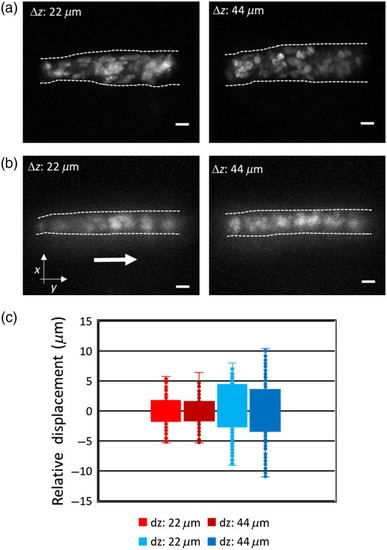
The maximum intensity projection of a volume image (10 slices) of RBCs in Tg(fli1a:EGFP; gata1:DsRed) zebrafish larva over two volume thicknesses using 2p LSFM (a) without and (b) with axicon imaging. (c) Boxplot of RBCs displacement in |
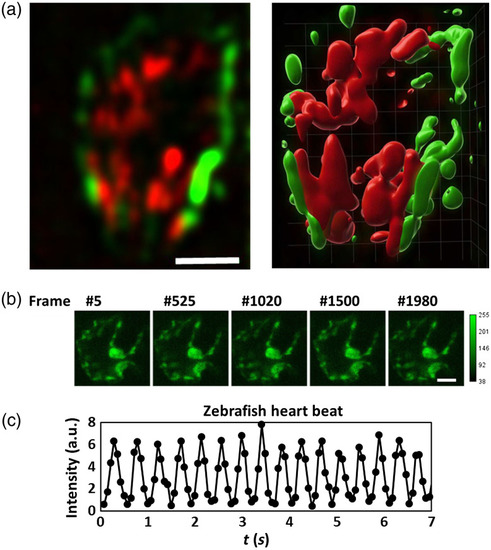
Two-color 2p LSFM with axicon imaging of a zebrafish heart beating. (a) A slice of a volume image and its 3D rendering image (10 slices; |