FIGURE SUMMARY
Title

The Role of Macrophages During Zebrafish Injury and Tissue Regeneration Under Infectious and Non-Infectious Conditions

Authors
Bohaud, C., Johansen, M.D., Jorgensen, C., Ipseiz, N., Kremer, L., Djouad, F.
Source
Full text @ Front Immunol

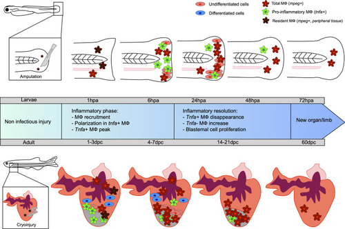
Kinetic of caudal fin and heart regeneration under non-infectious conditions. Transection of the zebrafish embryo caudal fin in non-infected condition leads to early recruitment of MФ, which complements the pool of resident MФ already present in the tail. At this very early stage, resident macrophages (MФ) phagocytose debris and dead cells. During the inflammatory phase, some MФ undergo polarization into tnfa-positive pro-inflammatory MФ, reaching a peak at 6 hpa. The tnfa-positive MФ disappear and a majority of non-inflammatory tnfa-negative MФ accumulate. During resolution of the inflammation, the undifferentiated blastema cells proliferate, peaking at 24 hpa, resulting in new limb formation. Cryoinjury of the heart of the adult zebrafish leads to an early recruitment of MФ, completing the pool of resident MФ already present in the heart, between 1 and 3 dpc. Tnfa-positive pro-inflammatory MФ predominantly arrive at the site and the proliferation of undifferentiated cells around the wound constituting the blastema begins. Already differentiated cardiomyocyte cells participate in tissue restoration. Non-inflammatory MФ represent the major cells present until the end of the regeneration process, resulting in a healed organ.

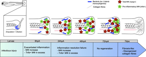
Caudal fin regeneration under infectious conditions. Amputation of the caudal fin in an infectious condition, with a scalpel pre-soaked in a solution containing pathogenic microorganisms (for instance Listeria monocytogenes) impairs tissue regeneration. Exacerbated inflammation is caused by an excessive number of tnfa-positive MФ at the wound. Resolution of inflammation is impeded at 24 hpa, with many tnfa-positive MФ that remain present. This results in incomplete tissue restoration and fibrosis at 7 dpa onward, characterized by the presence of disorganized collagen fibers.

Acknowledgments
This image is the copyrighted work of the attributed author or publisher, and ZFIN has permission only to display this image to its users. Additional permissions should be obtained from the applicable author or publisher of the image. Full text @ Front Immunol