
- Title
-
Advancing Diabetic Retinopathy Research: Analysis of the Neurovascular Unit in Zebrafish
- Authors
- Middel, C.S., Hammes, H.P., Kroll, J.
- Source
- Full text @ Cells

Anatomy of the zebrafish retina: Left: 4× magnification of a paraffin cut of the zebrafish retina, periodic-acid Schiff’s (PAS) stain. Right: 20× magnification of the zebrafish retina with a schematic overview of the different cell types. Scale bar = 100 μm. |
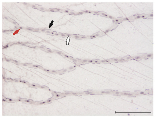
Visualization of endothelial cells, pericytes and erythrocytes in the zebrafish retina: 20× magnification of the zebrafish retina after digestion in 3% trypsin [81] and haemalum stain. Red arrow: erythrocyte, black arrow: pericyte, white arrow: endothelial cell. Scale bar = 100 μm. |