FIGURE SUMMARY
Title

Diesel exhaust exposure alters the expression of networks implicated in neurodegeneration in zebrafish brains

Authors
Jami, M.S., Murata, H., Barnhill, L.M., Li, S., Bronstein, J.M.
Source
Full text @ Cell Biol. Toxicol.

Visualization of expression analyses at proteome A and transcriptome B levels. A total of 11,172 proteins were identified from the TMT-labeled samples, among which 141 proteins were significantly upregulated and 607 downregulated A. The RNA-seq analysis detected 14,748 hits with 367 upregulated and 149 downregulated targets B

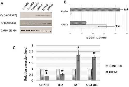
Validation of proteomic and transcriptomic findings. A and B Upregulation of Cyp1A protein and downregulation of Complexin 2 (CPLX2) was confirmed via western blot considering GAPDH as an internal control. C Higher levels of TAT and UGT1B1 transcription and lower levels of CHNRB and TH2 were also confirmed by qPCR using Elf-alpha as the internal control. The asterisks indicate statistical significance (∗ ∗  = p < 0.01, ∗  = p < 0.05)

The Metascape online tool allows for gene annotation for Danio rerio using combined proteomics and transcriptomics alterations. Several processes such as response to xenobiotic stimulus, metabolism of xenobiotics by cytochrome P450, and circadian regulation of gene expression were found induced upon DEPe treatment A, while “Visual perception,” “Phototransduction,” and “G protein-coupled receptor internalization” were suppressed B

Summarization of biological events during DEPe treatment. Red and green arrows indicate the alterations at protein and transcript levels, respectively

Cyp1A knockdown model. A Lack of induced expression of Cyp1A confirmed the KD efficiency; the expression levels were normalized to Elf-alpha as an internal control. Additionally, from the RNA-seq results table, the succinate dehydrogenase complex subunits B (SDHB) with stable expression between groups was selected as a secondary internal control. B The KD embryos had decreased survival in both treated and control conditions, but there was a significantly increased vulnerability to DEPe in KD embryos compared to controls. C Increased rate of morphological defects in KD embryos after exposure to DEPe demonstrates increased vulnerability in the absence of Cyp1A. The asterisks indicate statistical significance (∗ ∗  = p < 0.01, ∗  = p < 0.05)

Acknowledgments
This image is the copyrighted work of the attributed author or publisher, and ZFIN has permission only to display this image to its users. Additional permissions should be obtained from the applicable author or publisher of the image. Full text @ Cell Biol. Toxicol.