FIGURE SUMMARY
Title

A 127 kb truncating deletion of PGRMC1 is a novel cause of X-linked isolated paediatric cataract

Authors
Jones, J.L., Corbett, M.A., Yeaman, E., Zhao, D., Gecz, J., Gasperini, R.J., Charlesworth, J.C., Mackey, D.A., Elder, J.E., Craig, J.E., Burdon, K.P.
Source
Full text @ Eur. J. Hum. Genet.

Pedigree of family CRVEEH66 and candidate variant status.

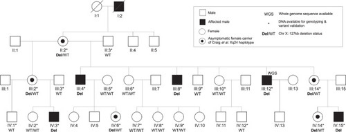
The previously studied CRVEEH66 family [12]. An ophthalmologist confirmed cataract affection status and no clinically significant lens opacity was observed in females. Segregation of 127 kb deletion (Del), at Xq24, is displayed. All affected males are hemizygous for the deletion and obligate female carriers are heterozygous for the variant (Del/WT) as is female IV:14.

CRVEEH66 genome-wide parametric linkage analysis results.

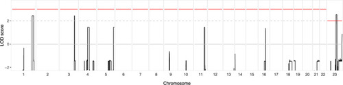
Multipoint LOD scores (
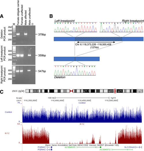
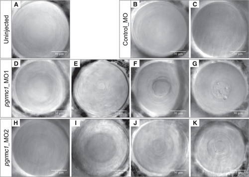
y-axis), indicating evidence for linkage, are displayed across the genome (x-axis). A linkage peak on chromosome X (Chr. 23), between markers rs2428312 and rs7887767, reaches significance with a maximum LOD score of 2.53. Regions on both chromosome 1 (rs696859–rs1316440) and chromosome 3 (rs4527385–rs4857688) reached maximum LOD scores of 2.44, with autosomal LOD scores exceeding two suggestive evidence of linkage (dashed line). The cut-off value for significance is indicated by the red line.

CRVEEH66 127 kb Xq24 deletion variant validation.

A Agarose gel electrophoresis results of representative samples for PCR products: deletion, left breakpoint, and right breakpoint. Heterozygous female obligate carrier and hemizygous affected male both possess the deletion amplicon. Unaffected female and male samples display wild-type results, with amplicons for breakpoints only. M; 100 bp ladder, C; PCR no template control. B Diagrammatic representation of the 127 kb deletion, indicated by the hatched region. Sequencing chromatograms at the critical breakpoints show consensus with the related halves of the deletion sequence. Deletion PCR fragment displayed from sequencing with the reverse primer. C UCSC genome browser displays the whole-genome sequence coverage from III:12 in dark red compared to an unrelated control sample in blue focused on the region surrounding the PGRMC1 gene. The coverage peaks within the deletion region (as indicated by the marked drop in coverage) in individual III:12 correspond with repeat regions in the genome (data not shown) and likely represent spurious mapping.

Zebrafish <italic>pgrmc1</italic> morpholino-induced knockdown lens phenotypes.

Representative DIC lens images from pgrmc1 morpholino experiments. Wild-type Gal4s1020t/UAS:mCherry transgenic zebrafish display healthy lenses (A). Control fish injected with standard control morpholino display healthy lens (B) and minor cataract with very fine nuclear pitting (C). Fish injected with pgrmc1_MO1 (2 ng) morpholino displayed both healthy lenses (D) or cataracts of variable severity; minor nuclear central mass (E), mild nuclear central mass with fibre cell disorganisation (F) and moderate/severe nuclear density with pitting (G). Fish injected with pgrmc1_MO2 (12 ng) morpholino displayed mainly healthy lenses (H) or subtle cataracts of variable severity; minor distinct central lens density (I), minor nuclear fibre cell disorganisation (J) and moderate nuclear fibre cell disorganisation with minor pitting (K).

ZFIN is incorporating published figure images and captions as part of an ongoing project. Figures from some publications have not yet been curated, or are not available for display because of copyright restrictions.

EXPRESSION / LABELING:
Gene:
Fish:
Anatomical Term:
Stage: Day 4
Acknowledgments
This image is the copyrighted work of the attributed author or publisher, and ZFIN has permission only to display this image to its users. Additional permissions should be obtained from the applicable author or publisher of the image. Full text @ Eur. J. Hum. Genet.