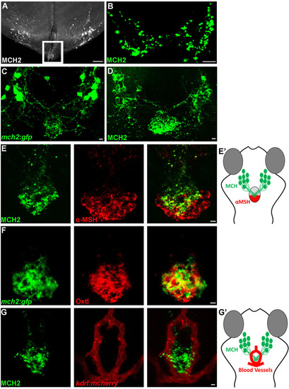
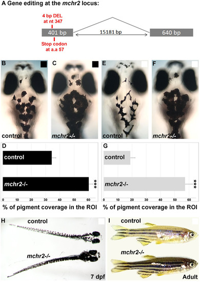
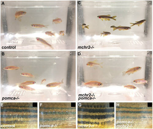
The hypothalamic MCH2 peptide interacts with the neurohypophysis.

(A-B) Confocal projection of immunolabelling with MCH in the adult zebrafish brain (A) and pituitary gland (B). The outline area represents the hypothalamic MCH projection at immediate vicinity of the adult zebrafish pituitary, lost during the tissue preparation. (C, D) Confocal projection of immunolabelling with EGFP in Tg(mch2:egfp) hypothalamus (C) or with the MCH2 antibody (D) at 5 dpf, reveals that MCH2 neurons in the hypothalamus have axonal projections on the pituitary gland. (E, E’) Confocal projection of double immunolabelling with MCH2 and α-MSH at 7 dpf, showing MCH cellular projections on the α-MSH+ domain (E). Schematic representation of the ventral part of the zebrafish brain showing the hypothalamic neurons expressing MCH2 targeting the posterior part of the pituitary gland expressing α-MSH (neurohypophysis) (E’). (F) Confocal projection of double immunolabelling with Oxtl and EGFP in Tg(mch2:egfp) hypothalamus at 7 dpf, demonstrating that MCH neurons target the neurohypophysis. (G, G’) Confocal projection of double immunolabelling with MCH2 and blood vessels at 5 dpf, suggesting that MCH peptide can be released in the bloodstream (G). Schematic representation of the ventral part of the zebrafish brain showing the hypothalamic MCH2 terminal projections targeting the neurohypophysis surrounded by blood vessels (Hypothalamo-Hypophyseal system) (G’). Ventral (A-C) or (D-F) dorsal view with anterior up. Scale bars: 50 μm (A) or 10 μm (B-G).

Mammalian orthologous MCH2 peptide induces melanosome contraction.

(A-C) Dorsal melanocytes of control (A), Tg(hs:mch1) (B) or Tg(hs:mch2) (C) black adapted larvae at 7 dpf following heat-shock. (D) Melanosome coverage was quantified in control, Tg(hs:mch1) or Tg(hs:mch2) in region of interest (ROI). The ROI has been defined as an area from the posterior of the eyes to the posterior of the hindbrain. Error bars represent s.d. *P<0.05, **P<0.001, ***P<0.0005, determined by t-test, two-tailed.

Loss of Mchr2-dependent signaling leads to melanosome dispersion.

(A) Schematic representation of the mchr2 locus and the small genomic deletion induced by CRISPR/Cas-9 leading to a premature stop codon in the mchr2 coding sequence. (B, C) Dorsal melanocytes of control (B) or mchr2 homozygous mutant (C) black adapted larvae at 7 dpf. (D) Melanosome coverage was quantified in control or mchr2 homozygous mutant black adapted larvae at 7 dpf in ROI, an area from the posterior of the eyes to the posterior of the hindbrain. (E, F) Dorsal melanocytes of control (E) or mchr2 homozygous mutant (F) white adapted larvae at 7 dpf. (G) Melanosome coverage was quantified in control or mchr2 homozygous mutant white adapted larvae at 7 dpf. (H, I) Larval and adult mchr2 homozygous mutant phenotypes are characterized by melanosome dispersion on a white background, indicating that MCH signaling is required to promote melanosome contraction in melanocytes. Dorsal view with anterior up (B, C, E, F and H). Lateral view (I). Error bars represent s.d. *P<0.05, **P<0.001, ***P<0.0005, determined by t-test, two-tailed.

MCH-dependent melanosome contraction relies on Mchr2 activity.

(A-D) Dorsal melanocytes of control (A), mchr2 homozygous mutant (B), mchr2 homozygous mutant; Tg(hs:mch1) (C) or mchr2 homozygous mutant; Tg(hs:mch2) (D) black adapted larvae at 7 dpf following heat-shock. (E) Melanosome coverage was quantified in control, mchr2 homozygous mutant, mchr2 homozygous mutant; Tg(hs:mch1) or mchr2 homozygous mutant; Tg(hs:mch2) larvae at 7 dpf. Dorsal view with anterior up. Error bars represent s.d. *P<0.05, **P<0.001, ***P<0.0005, determined by t-test, two-tailed.

<italic>pomca</italic> loss of function leads to melanosomes contraction, but is not sufficient to rescue the <italic>mchr2</italic> mutant phenotype in larvae.

(A) Schematic representation of the pomca locus and the small genomic deletions induced by CRISPR/Cas-9 leading to premature stop codons in the pomca coding sequence. (B-C’) Confocal projection (B, C) or section (B’, C’) of double immunolabelling with MCH2 and α-MSH antibody at 5 dpf, showing that MCH expression, but not α-MSH, is detected in the pomca homozygous mutant. (D, E) Dorsal melanocytes of control (D) or pomca homozygous mutant (E) black adapted larvae at 7 dpf. (F) Melanosome coverage was quantified in control or pomca homozygous mutant black adapted larvae at 7 dpf. (G-J) Dorsal melanocytes of control (G), pomca homozygous mutant (H), mchr2 homozygous mutant (H) or pomca; mchr2 double homozygous mutant (J) white adapted larvae at 7 dpf. (K) Melanosome coverage was quantified in control, pomca homozygous mutant, mchr2 homozygous mutant or pomca; mchr2 double homozygous mutant white adapted larvae at 7 dpf. Dorsal view with anterior up (D, E and G-J). Scale bars: 10 μm. Error bars represent s.d. *P<0.05, **P<0.001, ***P<0.0005, determined by t-test, two-tailed.

MCH signaling counteracts melanocortin system activity to promote melanosome contraction in adult zebrafish.

(A-D) Loss of function phenotype on skin pigmentation from white adapted adult control (A), pomca(B), mchr2(C) or double mchr2; pomca(D) homozygous mutant. (E-G) Loss of function phenotype on skin pigmentation from black adapted adult control (E), pomca(F), mchr2(G) or double mchr2; pomca(H) homozygous mutant. Similar to the pomca homozygous mutant, pomca; mchr2 double homozygous mutant phenotype is characterized by melanosome contraction on a black background, indicating that pomca loss of function is sufficient to allow melanosome contraction in absence of MCH signaling in adult zebrafish. Lateral view (E-G).

Acknowledgments
This image is the copyrighted work of the attributed author or publisher, and ZFIN has permission only to display this image to its users. Additional permissions should be obtained from the applicable author or publisher of the image. Full text @ PLoS Genet.