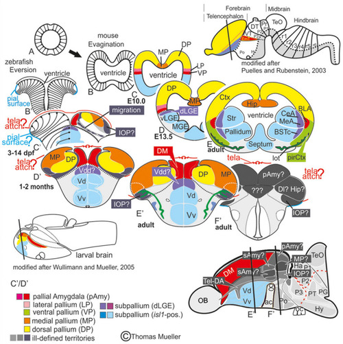
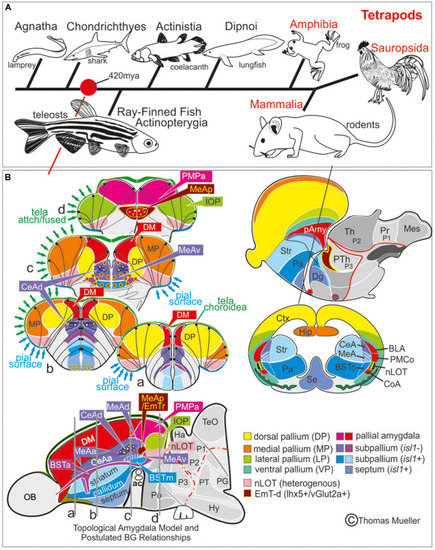
Telencephalic eversion in zebrafish and comparison to mammals. The schematic illustrates how both the outward-growing (eversion) process of the developing telencephalon and its adult morphology of zebrafish (lower row) compares to the telencephalon development (evagination) of mammals (upper row). (A) The telencephalon develops from the anteriormost part of the neural tube. (B–E) In mammals, two bilateral hemispheres develop around a centrally located ventricle. Predominantly glutamatergic pallial zones (warm colors) develop in the dorsal telencephalon, whereas mostly GABAergic subpallial territories (cold colors) are found in the ventral telencephalon. (B’–E’) Likewise, in teleosts like zebrafish, the dorsal and ventral telencephalon, respectively, hold pallial and subpallial territories, however, the ventricle due to the eversion comes to lie on top of the brain. (C’,D’) Proliferation patterns, BrdU-long term labeling, and gene expression studies in zebrafish indicated a complex eversion process that includes a radial migration toward the dorsoposterior pallial (“Dp”) zone (Mueller et al., 2011). The zebrafish dorsal pallium (yellow) subsequently gets overgrown by the pallial amygdala and the medial pallium (MP in orange). (F’) The teleostean eversion process is not comprehensively understood. A major obstacle in the comprehensively understanding both the eversion and comparative anatomy remains the unsolved delineation of the integrative olfactory pallium (IOP – the putative homolog to the mammalian piriform cortex) and of telencephalic entities in the posteriormost extent. Ambiguous structures and focus of this study are colored in gray.

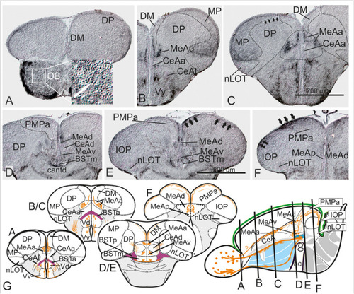
Molecular Definitions of the Zebrafish Amygdaloid Complex. Precommissural (A–E), supra- and postcommissural (F–Q) telencephalon, and delineation of the dorsal pallium in its periventricular {(S+T, U1/left) and central (U1, right, U2)} portions. Pallium: Analyzing the distributions of GABA (A–C,H,K), parvalbumin (E,F,J,N), and GFP in the transgenic line Tg(vGlut2a:GFP)(A–C,H,I,K,M) allowed to discern the dorsomedial pallial zone, the putative core region of the pallial amygdala, from the hippocampal division (medial pallium/MP) and the teleostean dorsal pallium (DP) (A–C). DP lacks both vGlut2a-driven GFP and parvalbumin-positive neurons but shows parvalbumin positive fibers (F,J,H,U2). The strongly GFP-positive DM lacks parvalbumin expression (F,J). The mostly vGlut2a:GFP-free hippocampal division (MP) exhibits both parvalbumin-positive fibers and neurons in addition to GABA-neurons (C,F,J). Only at posteriormost sections, we found vGlut2a:GFP positive neurons in the MP (H). At anteriormost sections (A,B), the DP reaches the dorsalmost periventricular zone, which holds proliferative stem cells. More posteriorly (C,F,J), the DP is shown in its secondarily centralized position overgrown by both the pAmy and hippocampal division (medial pallium/MP). In addition, we found many large GABAergic interneurons in DP (A–C), as well as sparsely distributed GABAergic interneurons in the pAmy and MP (B,C). Subpallium: Dense population of GABAergic neurons define the subpallium (A–C,H,K). Within the subpallium, isl1-driven GFP labels striatopallidal and septal divisions as well as the BSTm (D1,D2,F,G). In contrast, GABAergic territories that lack isl1-driven GFP define the majority of subpallial amygdaloid territories. The anterior (precommissural) dorsalmost. GABAergic population (CeAa) together with the migrated GABAergic nuclei (CeAl = former Vc) predominantly form the zebrafish central amygdala [CeA in (A–C)]. The ventricular-close GABAergic territory is juxtaposed and contiguous with the former supracommissural (Vs) and postcommissural (Vp) nuclei of medial amygdala (MeA; former Vs/Vp). In addition, based on the lack of isl1-driven GFP in both anterior and posterior TH-positive dopaminergic and their previous reported projection to the hypothalamus, we consider as the anterior and posterior bed nucleus of the stria termalis (BSTa/BSTpd). The medial bed nucleus of the stria terminalis (BSTm) is the only isl1:GFP positive medial nucleus [BSTm in (F,G)]. We also found sparse dopaminergic neurons within the posteromedial amygdala (MeAp) that project into part of the nLOT (B2). Supra- and postcommissural extended medial amygdaloid nuclei (MeAc, MeAd, MeAp, BSTm, BSTpd) are defined by the presence of numerous calretinin-positive neurons (F,I,M) similar to the mammalian situation. In contrast, the CeAd that comprises numerous laterally displaced GABAergic neurons shows many parvalbumin-positive fibers and few parvalbumin neurons, but lacks calretinin neurons. The MeAp was formerly assigned to the teleostean subpallium, because there are GABAergic neurons in its vicinity (K). However, the otp-a (L) and calretinin (I,M) positive neurons that define the MeAp are likely glutamatergic and originate from the thalamic eminence according to our results. Discerning PMPa, IOP, and nLOT Comparing secondary olfactory projections in the transgenic line Tg(lhx2a:GAP-YFP)(J,N) with parvalbumin allowed us to identify primary olfactory pallial territories and their topological relationships. The distribution of secondary olfactory projections in comparison to parvalbumin and calretinin fibers identifies two olfactory integrative structures: (1) the parvalbumin-positive integrative olfactory pallium (IOP) (I,J,M,N) formerly misinterpreted as part of the medial pallium (“Dl/Dlp”) in zebrafish; (2) the largely parvalbumin-free (migrated) amygdaloid nucleus of the lateral olfactory tract (nLOT). The nLOT is a predominately glutamatergic nucleus that consists of both vGlut2a:GFP positive (anterior; C) and negative (posterior) domains as well as GAD67-positive [arrows in (O,P)] and gad1b-driven dsRed expressing [arrow in (Q)] GABAergic zones resembling the mammalian nLOT that consists of alternating GABAergic and glutamatergic layers. Previously, the nLOT has been mislabeled as the posterior division of the dorsal telencephalon (“Dp”) and mistakenly considered the piriform cortex homolog. The identification of the nLOT together with the piriform cortex homolog (IOP) solves their topological relationships within the complexly everted telencephalon (R indicates orientation of sections A–N). (F) Schematic figure shows orientation of section in the sagittal view. New hallmarks of the complexly everted zebrafish telencephalon – the rostral (superficial) versus posterior centralized parts of the dorsal pallium (DP). (S–U2) Analyzing distribution of parvalbumin versus vGlut2a-driven GFP shows that the dorsal pallium reaches the periventricular zone and includes its germinative layer of origin. Note, that DM and medial pallium (MP = dorsolateral (Dl) zone of the pallium) overgrow the dorsal pallium at posterior sections at the point of convergence (circled area). (V) Schematics of a zebrafish telencephalon from dorsal perspective illustrating levels of cross sections.

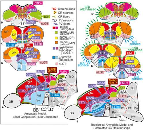
Molecular code of the zebrafish amygdaloid complex. Definitions of amygdaloid territories in the complexly everted telencephalon: Amygdala model (basal ganglia (BG) not considered) (A–D) versus idealized topological amygdala model (B’,C’,D’,E,F) indicating tela attachments and radial glia distribution. (A–D) The lateral schematic (A) of the amygdala model divides the zebrafish amygdaloid complex with regard to the anterior commissure (ac) into precommissural (B), supracommissural (C), and postcommissural (D) sectors. A hierarchical code defines all amygdaloid nuclei: All medial extended amygdaloid nuclei in the supra- and postcommissural sectors (C,D) are located within the isl1:GFP negative, subpallial (GABAergic) territories and comprise numerous calretinin-positive neurons. The CeAd is distinguished from such MeA-territories through the presence of parvalbumin-fibers and cells that are laterally displaced. More anterior lying CeA-territories such as the CeAa, CeAl also form part of the isl1:GFP negative subpallium, they are, however, distinguished from the CeAd through their lack of parvalbumin expression. In addition, all dopaminergic clusters formerly viewed to the zebrafish striatopallidum are here considered the anterior (BSTa) and posterior (BSTp) divisions of the bed nucleus of the stria terminalis (BST). These dopaminergic extended amygdaloid nuclei are also located in the zebrafish isl1:GFP free territory that corresponds to the mammalian dLGE (= Vdd). The medial BST(m) is the only subpallial amygdaloid territory where a large population of isl1:GFP forms the majority of this nucleus in addition to some calretinin-positive neurons that link this nucleus with the rest of the extended medial amygdala. The newly discovered integrative olfactory nucleus (IOP) shows secondary olfactory projections and many parvalbumin positive neurons. In contrast, the amygdaloid nLOT that also receives secondary olfactory projections lacks these parvalbumin neurons. (E) Topological amygdala model indicating idealized BG relationships showing tela attachment sites and radial glia distribution as revealed in this study.

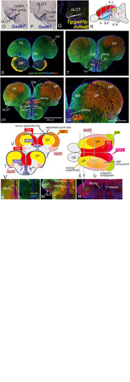
Substance P Fiber Tracts from the Olfactory Bulb into the Telencephalon. (A–G) Using diaminobenzidine (DAB) as a substrate for the detection of a horseradish peroxidase coupled antibody yielded enhanced detection of substance P in the telencephalon (A–G). We found substance P positive neurons in the olfactory bulb (OB in A and inlet). substance P fibers emanate from the olfactory bulb toward the lateral ventralmost part of the subpallium [Vv+Vd in (B,C)]. Most importantly, some SP cells and many SP fibers label the medial amygdala (MeAa) in the dorsal tier of the subpallium (B–F). At the transition between supracommissural and postcommissural telencephalon (D–F), substance P positive fibers passing through the dorsal medial amygdala [MeAd in (D–F)] enter and label the posteromedial pallial amygdala [PMPa in (E,F)]. Substance P fibers also pass through the PMCo into the IOP, the zebrafish putative piriform cortex homology (black arrows in IOP of H), supporting the proposed homology between the zebrafish PMPa and mammalian PMCo. Note, the anterior and dorsoposterior divisions of the CeA (CeAa + CeAd) lack substance P and therefore can easily distinguished from all medial amygdaloid nuclei (MeAa, MeAd, MeAv, MeAp). (G) Schematic cross sections and sagittal view of the zebrafish telencephalon to illustrate the two solid zebrafish accessory olfactory substance P fiber systems innervating the medial amygdaloid territories. Substance P fibers seemingly form also laterally displaced diffuse projections (E).

Radial glia cells and Tela Attachement Sites Confirm Medial, Dorsal, and Thalamic Eminence Identification. We performed triple fluorescence immunostains against parvalbumin (PV), GFP, and GFAP in the brains of Tg(vGlut2a:GFP) counterstained against DAPI. (A1–C2): A1 shows different aspects of a rostral section highlighting that one large parvalbumin- (PV-) positive tela attachment site is located at the ventricular site of the dorsal pallium and pallial amygdala (white arrows in A1,A2,B,C1). The tela choroidea is fused (green arrows in A1,A2,B,C1) with the proliferative stem cell layer of the dorsolateral zone (“Dl”) supporting its medial pallial (“hippocampal”) identity. The nLOT tract shows a characteristic indentation (A2,C2) and no GFAP stained cell somata at its lateralmost aspect corroborating this area as the pial, not ventricular site. Notably, all radial glia of the candidate dorsal pallium do send their processes towards the pallial-subpallial border (PBS). These radial glia are attached to the topographically ventral side of the nLOT next to those of the medial pallium. Radial glia of the nLOT are not visible in this orientation suggesting that their radial domain is different from pallial zones. This finding supports the interpretation that the vGlut2a-driven GFP domain is a derivative of the EmT. (D1A–D2B) At mid-telencephalic sections, both radial glia cells and tela attachments show the same distribution. At the point where proliferative zones of the DM and medial pallium (MP) meet and grow over the dorsal pallium, a reduced number of radial glia cells of the dorsal pallium are visible. Radial glia of both MP and DM cut through the territory of DP and send their processes beneath the nLOT. The probable diencephalic attachment site of the tela choroidea is located at the nLOT (gray arrows in D1b,D2b). Again, there are no GFAP-positive glia cell somata at the most lateral cell groups corroborating its pial nature. The nLOT in this orientation shows no radial glia processes supporting their postulated EmT origin. At caudalmost sections, the study identifies the diencephalic -telencephalic junction (DTJ) that comprises territories formerly assigned to the telencephalon such as the posterior medial amygdala (MeAp/EmTr) and the vGlut2a-driven GFP positive nLOT territory that spans a region next to the MeAp/EmTr to the lateral (pial) surface (blue arrows in E1-E3). The BNSM was formerly identified as a derivative of the EmT in zebrafish and is often mistaken as the entopeduncular nuleus proper (ENv) Mueller and Guo 2009). Note, that the tela choroidea is attached to the MeAp/EmTr indicating its periventricular site (E1,F1).

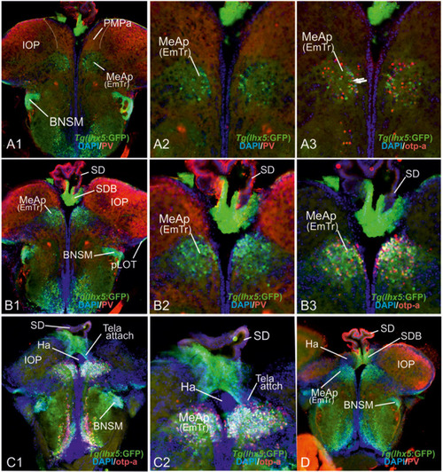
The posterior medial amygdala (MeAp) is identical with the rostral thalamic eminence (EmTr). (A1–D): The comparison between lhx5-driven GFP, parvalbumin (PV), and otp-a protein distribution shows that the lhx5-driven GFP positive EmTr reaches from its periventricular site (indicated by tela choroidea attachment in C). Note, the lhx5-driven GFP positive base (SDB) of the saccus dorsalis (SD) connects to both the Ha (in C1/2 and D) and the MeAp/EmTr (best visible in C1/2)hh. Like in mice, the posterior territory of the lateral olfactory tract seemingly does express lhx5 as indicated by GFP expression in Tg(lhx5:GFP) (A1,B1,C1,D).

Comparison Zebrafish – Macrosmatic Rodent Amygdala. (A) Cladogram indicating presence of a complex amygdala ground plan and nucleus of the lateral olfactory tract (nLOT) in the last common ancestor of teleosts and mammals. Our results indicate that the last common ancestor between ray-finned fish and mammals already showed a tetrapod-like main extended amygdala ground plan (red circle). Previously, most scientists assumed that a bipartite main versus olfactory extended amygdala evolved with a vomeronasal epithelium in the last common ancestor of lungfish and tetrapods (González et al., 2010). However, molecular evidence hint toward the presence of a bipartite olfactory system already in agnathan lamprey (Chang et al., 2013). We speculate, therefore, that a bipartite and complex amygdala may evolved with the earliest vertebrates. (B) Prosomeric comparisons of the zebrafish/teleostean amygdala (left) with the situation in macrosmatic rodents (right). Both the schematized zebrafish brain cross sections and the parasagittal view on the left side indicate that the zebrafish amygdala holds several previously misinterpreted territories such as the bed nucleus of the stria terminals (BST), the medial amygdala (MeA) and its anterior (MeAa), posterior (MeAv) and dorsal division (MeAd), the posteriormedial pallial amygdala (PMPa), the integrative olfactory pallium (IOP), and the nucleus of the lateral olfactory tract (nLOT). Due to the teleostean-specific outward growing process (eversion), these territories lie on top of the telencephalon and cover the zebrafish homolog to the mammalian isocortex. In mammals (right side), we find the opposite situation. Here, the amygdaloid complex is located in the anterior ventral depth of the brain covered by the enlarged isocortex (modified after Mueller et al., 2011; Mueller, 2012). For molecular definition of pallial, subpallial, and EmT-derived (EmT-d) amygdaloid territories see Table 1. Abbreviations see table.

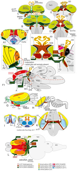
Summary schematic explaining how the zebrafish thalamiceminence (EmT) reveals ancestral topological relationships to posterior medial amygdala and olfactory pallium. (A–F2) Comparing topographically rostral and posterior ends of the telencephalon in transgenic lines Tg(vGlut2a:GFP) and Tg(lhx5:GFP) with distribution of parvalbumin, otp-a, and GFAP indicated a complex EmT. In sites classically defined as “telencephalic,” we identified the rostral (EmTr = vGlut2a+, lhx5+, otp-a+) and lateral (EmTl) vlgut2a+;lhx5-). At diencephalic side of the diencephalic-telencephalic junction, the EmTm can be easily identified based on vGlut2a-driven GFP expression and absence of lhx5-driven GFP, whereas the otp-a negative bed nucleus of the stria medullaris (BNSM) shows both vlgut2a- and lhx5-driven GFP. Note, that we consider the rostral and posteriormost parts of the newly defined nLOT a derivative of the EmT consistent with the absence of radial glia somata and processes in their most lateral (pial) aspects. (G–L) Schematics comparing brain of adult zebrafish (dorsal view) with adult rodent (idealized side view), and developmental stage to determine various EmT derivatives. The study identifies topographically rostral and posterior EmT territories based on differential expression of vGluta- and lhx5-driven GFP in comparison to otp-a expression. The dorsal view on the zebrafish telencephalon highlights “diencephalic” medial (EmTm) versus “telencephalic” rostral (EmTr) and lateral (EmTl) at the diencephalic-telencephalic junction (DTJ). The distribution of vGlut2a-driven GFP and lhx5-driven GFP in adult zebrafish strikingly resembles the situation shown in mammals (mouse) (Ruiz-Reig et al., 2017). Our complexly everted telencephalon model considers both rostral (nLOTr) and posterior portions of the most ventro-lateral expression domains of vGlut2a-driven GFP as radial extensions of the EmTl. The integrative olfactory pallium (IOP) is most likely a derivative of the newly defined lateral pallium of the prosomeric model and thus homologous to the entorhinal cortex.

Acknowledgments
This image is the copyrighted work of the attributed author or publisher, and ZFIN has permission only to display this image to its users. Additional permissions should be obtained from the applicable author or publisher of the image. Full text @ Front. Neurosci.