FIGURE SUMMARY
Title

Inhibition of ZIP4 reverses epithelial-to-mesenchymal transition and enhances the radiosensitivity in human nasopharyngeal carcinoma cells

Authors
Zeng, Q., Liu, Y.M., Liu, J., Han, J., Guo, J.X., Lu, S., Huang, X.M., Yi, P., Lang, J.Y., Zhang, P., Wang, C.T.
Source
Full text @ Cell Death Dis.

Association of ZIP4 expression with clinicopathological variables in patients with NPC.

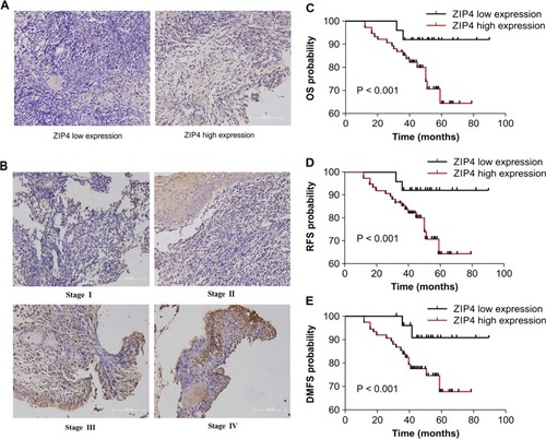
Representative specimens from patients with NPC showed weak (a) and strong (b) cell membrane immunostaining for ZIP4 at different NPC stages (Scale bar = 101 μm). The results of the Kaplan–Meier survival analysis indicate that patients with high ZIP4 expression present a shorter overall survival (OS) (c), recurrence-free survival (RFS) (d), and distant metastasis-free survival (DMFS) (e). The differences in OS, RFS, and DMFS between the ZIP4 low and high expression levels were significant, low/high = 25/74 (P < 0.001)

ZIP4 silencing inhibited migration and invasion capabilities of C666-1 cells.

a Relative protein and mRNA level of ZIP4 were detected in stable LVRH, Sh-ZIP4, Control, and ZIP4 cell lines. b The cell migration rate between LVRH C666-1 and Sh-ZIP4 C666-1 cells was compared by the wound-healing assay. Microscopic observation was recorded at 0 and 24 h after scratching the cell layer (Scale bar = 50 μm). c The invasive and migration properties of the cells were analyzed by using a matrigel-coated Boyden chamber. Migrated cells were plotted as the average number of cells per field from three different experiments (Scale bar = 50 μm). d The expression levels of EMT markers, E-cadherin (E-cad), vimentin (Vim), and FSP-1 were examined by immunofluorescence staining (Scale bar = 75 μm). e E-cad, vimentin (Vim), and FSP-1 expression were examined in control and ZIP4 C666-1 cells by western blot. The experiments were performed in triplicate. The data are represented as means ± SD from three independent experiments. *P < 0.05

ZIP4 silencing prevents C666-1 tumor cell invasion, dissemination, and metastasis in a zebrafish model and a lung metastasis mouse model.

a, b Red fluorescence-labeled tumor cells were injected into the perivitelline space of 48 h postfertilization embryos and tumor cell invasion, dissemination, and metastasis were detected using confocal microscopy at day 3 post injection. Yellow arrows indicate primary tumors. White arrows indicate disseminated tumor foci (Scale bar = 100 μm). c Quantification of disseminated tumor foci (n = 40 per group), P < 0.01. d Representative images of the mouse lungs. e The average number of tumor metastatic nodules in the lungs of mice xenografted with C666-1, LVRH C666-1 cells, and Sh-ZIP4 C666-1 cells is provided (N = 7; P < 0.01). f Representative views of lung tissue sections from each group are shown (Scale bar = 50 μm). The experiments were performed in triplicate. The data are represented as means ± SD from three independent experiments

ZIP4 associated signaling pathways and mechanism were enriched.

a KEGG pathway analysis of the discrepantly expressed genes in Sh-ZIP4 compared with control LVRH C666-1 cells. b Phospho-antibody array. See Supplementary Fig. 1 for full results and additional annotations. c The western blot assay of the selected proteins for the determination of phosphorylation antibody array. d Effect of PI3K inhibitors, LY294002, and Wortmannin on EMT markers. The experiments were performed in triplicate. The data are represented as means ± SD from three independent experiments. *P < 0.05

ZIP4 silencing enhances radiosensitization in vitro.

a Cell growth in low and high concentration of zinc. b Inhibition of cell proliferation following exposure to various doses of Co60 was evaluated by the MTT assay (P < 0.01). c Images and quantification of the number of colonies formed from cells treated with 0, 2, 4, 6, and 8 Gy (P < 0.05). d Flow cytometry analysis of apoptosis using annexin V and propidium iodide in cells treated with various doses of radiation (0, 2, 4, 6, and 8 Gy). e Flow cytometric analysis of the cell cycle in cells treated with various doses of radiation. f Protein expression levels of cleaved caspase-3, cleaved caspase-9, Bax, and Bcl-2 in cells treated with a 6 Gy dose of radiation. Experiments were repeated three times. The data are represented as means ± SD from three independent experiments. *P < 0.05

ZIP4 silencing enhances the antitumor effect of radiation in vivo and Schematic summary of this study.

a, b Sh-ZIP4 and LVRH C666-1 cells (1 × 107) were subcutaneously inoculated into the right hind limb of nude mice (N = 7 per group). When the size of the tumors reached about 50 mm3, tumors were irradiated at 5 Gy per day for 4 days (on day 1, 3, 6, and 8). Tumor size was measured and weight was recorded. c TUNEL staining of C666-1 xenografts (Scale bar = 50 μm) (P < 0.01). d PCNA IHC staining of C666-1 xenografts (P < 0.01). e Overexpression of ZIP4 induces the EMT process through the PI3K/Akt pathway and further promotes migration and invasion of NPC cells. Meanwhile, ZIP4 enhanced radiation-induced apoptosis and growth inhibition of human C666-1 cells. These findings support our clinical observations, indicating that ZIP4 elevation is associated with clinical T classification, N classification, and clinical stage, and leads to adverse survival outcomes of NPC

Acknowledgments
This image is the copyrighted work of the attributed author or publisher, and ZFIN has permission only to display this image to its users. Additional permissions should be obtained from the applicable author or publisher of the image. Full text @ Cell Death Dis.