
- Title
-
PRL3 enhances T-cell acute lymphoblastic leukemia growth through suppressing T-cell signaling pathways and apoptosis
- Authors
- Garcia, E.G., Veloso, A., Oliveira, M.L., Allen, J.R., Loontiens, S., Brunson, D., Do, D., Yan, C., Morris, R., Iyer, S., Garcia, S.P., Iftimia, N., Van Loocke, W., Matthijssens, F., McCarthy, K., Barata, J.T., Speleman, F., Taghon, T., Gutierrez, A., Van Vlierberghe, P., Haas, W., Blackburn, J.S., Langenau, D.M.
- Source
- Full text @ Leukemia
|
|
|
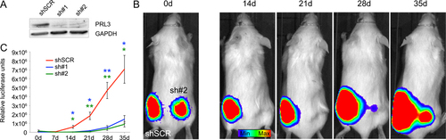
(A) Western blot analysis following shRNA knockdown in Jurkat cells. (B) Luciferase bioluminescent imaging of representative animal engrafted with scramble control shRNA (shSCR, left flank) compared with shRNA to PRL3 (sh#2, right flank). High exposure images shown on 0 day to ensure equal injection of control and knockdown cells (left panels). Animals shown at different exposure from 14–35 days to highlight relative differences in growth between control and knockdown cells (right panels). (C) Quantification of growth at different time points. * denotes p<0.05, ** denotes p<0.01, Student’s t-test. Error bars denote standard error of the mean. N ≥ 5 mice/experimental arm. |
|
|
|