
- Title
-
Cardioprotective Effects of MTSS1 Enhancer Variants
- Authors
- Morley, M.P., Wang, X., Hu, R., Brandimarto, J., Tucker, N.R., Felix, J.F., Smith, N.L., van der Harst, P., Ellinor, P.T., Margulies, K.B., Musunuru, K., Cappola, T.P.
- Source
- Full text @ Circulation
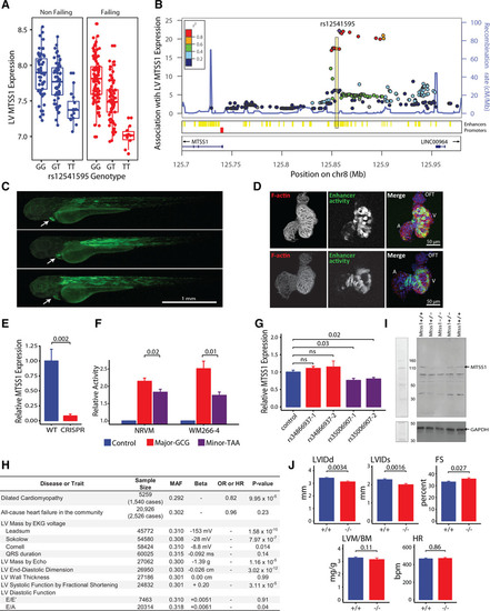
Interrogation of the MTSS1 locus.A, Association of minor allele (T) at rs12541595 with MTSS1 expression in human left ventricle (LV; P=6×10–23 across all samples using an additive genetic model adjusting for presence/absence of heart failure; n=313). B, cis associations with LV MTSS1 expression (–log10P values using same model as in A; n=313). Variants within a putative enhancer (yellow) in linkage disequilibrium with rs12541595 show strong associations. C, The enhancer region was cloned upstream of a green fluorescent protein reporter and transfected into zebrafish embryos to determine the anatomic site of activity (green). Activity is specific to cardiac muscle (white arrow) and skeletal muscle (dorsal axis) 72 hours postfertilization. D, Analysis of cardiac-restricted enhancer activity within excised zebrafish hearts demonstrates strong activity (green) in the ventricle (V) and limited activity in the atrium (A) and outflow tract (OFT). Hearts are counterstained for sarcomeric architecture (F-actin, red) and nuclei (blue). E, CRISPR-Cas9 enhancer deletion abolishes MTSS1 expression in WM266-4 cells (n=6/group; Mann-Whitney U test P value). F, Relative activity of major and minor enhancer haplotypes defined by rs12541595, rs35006907, and rs34866937. The minor enhancer haplotype, marked by the rs12541595 minor allele, shows reduced activity (n=6/group; Mann-Whitney U test P values). G, CRISPR interference targeting enhancer variants shows reduced activity with dCas9 positioned at rs35006907 using 2 different guide RNAs (n=3/group; Mann-Whitney U test P values). H, Association of the MTSS1 rs12541595 minor allele (T) with cardiac traits in human populations (P values by additive genetic models). I, immunoblot of murine myocardial protein extracts verifying successful knock-out of Mtss1. J, LV structure and function in Mtss1+/+ (n= 23; 8 female, 15 male) and Mtss1–/– (n=16; 10 female, 6 male) mice by echocardiography under isoflurane anesthesia (gender-adjusted P values). FS indicates fractional shortening; HR, heart rate; LVIDd, LV internal dimension in diastole; LVIDs, LV internal dimension in systole; LVM/BM, LV mass normalized to body mass; MAF, minor allele frequency; NRVM, neonatal rat ventricular myocytes; ns, not significant; OR, odds ratio; and WT, wild type. |