FIGURE SUMMARY
Title

Octominin: A Novel Synthetic Anticandidal Peptide Derived from Defense Protein of Octopus minor

Authors
Nikapitiya, C., Dananjaya, S.H.S., Chandrarathna, H.P.S.U., De Zoysa, M., Whang, I.
Source
Full text @ Mar. Drugs

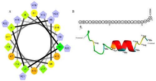
Predicted helical secondary and three-dimensional structures of Octominin. (A) Helical wheel of Octominin shows the AAs arrangement and the residue numbers which are counted from the amino (N) terminal of the peptide. The hydrophilic and hydrophobic residues are represented by circles and diamonds, respectively. The potentially charged (positively charged) residues are marked as pentagons in light blue. The most hydrophobic residue is green, and the amount of green is decreasing proportionally to the hydrophobicity, with zero hydrophobicity coded as yellow. (B) Three-dimensional structure and AA sequence of Octominin with positively charged residues.

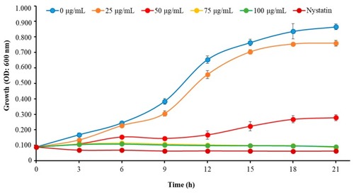
Time–kill kinetics of Octominin against C. albicans. C. albicans growth was assessed after Octominin treatment (0, 25, 50, 75, and 100 μg/mL) at 3 h intervals by measuring the optical density (OD) at 600 nm. The bars indicate the mean ± standard deviation (n = 3).

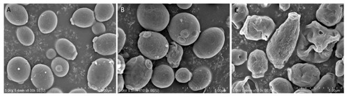
Effect of Octominin on morphological and structural changes of C. albicans assessed by field-emission scanning electron microscopy (FE-SEM). (A) Untreated C. albicans; (B) treated with 50 µg/mL (minimum inhibitory concentration; MIC;) Octominin; (C) treated with 200 µg/mL (minimum fungicidal concentration; MFC) Octominin.

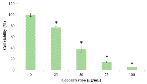
Effect of Octominin on the viability of C. albicans. Cell viability was assessed by the 3-(4,5-dimethylthiazol-2-yl)-2,5-diphenyltetrazolium bromide (MTT) assay after treatment with different concentrations of Octominin (0–100 μg/mL) until 24 h. * P < 0.05 compared to the control (untreated) group. The error bars indicate the mean ± standard deviation (n = 3).

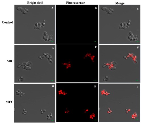
Effect of Octominin on membrane permeability in C. albicans. Confocal laser-scanning microscope (CLSM) merged and fluorescence images representing the cell membrane permeability by propidium iodide (PI) staining in C. albicans cells treated with Octominin at 30 °C for 6 h. (AC) Untreated control; (DF) treatment with Octominin at the MIC (50 µg/mL); (GI) treatment with Octominin at the MFC (200 µg/mL).

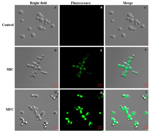
Effect of Octominin on reactive oxygen species (ROS) production of C. albicans. CLSM merged and fluorescence images representing ROS production in C. albicans with Octominin treatment at the MIC and MFC. (AC) Untreated control; (DF) treatment with Octominin at the MIC (50 µg/mL); (GI) treatment with Octominin at the MFC (200 µg/mL).

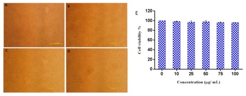
Cytotoxic effect of Octominin on HEK293 cells. (
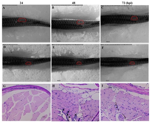
AD) Representative images of Octominin-treated HEK293 cells at 48 h post treatment. (A) Untreated (control), (B) 10 µg/mL Octominin, (C) 75 µg/mL Octominin, (D) 100 µg/mL of Octominin. (E) Cell viability percentage of HEK293 cells treated with Octominin at different concentrations (0, 10, 25, 50, 75, and 100 µg/mL). Data are expressed as the mean ± standard error (n = 3).

Anticandidal effect of Octominin in C. albicans-infected zebrafish. C. albicans growth and wound area at 24, 48, and 72 h post treatment of Octominin compared with the control (water-treated) fish. (AC) C. albicans infected nontreated control. (DF) C. albicans-infected Octominin treated fish. Effect of Octominin treatment in zebrafish muscle tissue upon C. albicans infection evaluated by periodic acid-Schiff-hematoxylin (PAS-H)-stained sections under microscope (400×). Muscle tissues of uninfected (G), infected and untreated treated (H), and infected and Octominin-treated (I) zebrafish. Black arrowhead shows the leukocyte infiltration in the muscle tissue of zebrafish.

Acknowledgments
This image is the copyrighted work of the attributed author or publisher, and ZFIN has permission only to display this image to its users. Additional permissions should be obtained from the applicable author or publisher of the image. Full text @ Mar. Drugs