FIGURE SUMMARY
Title

Parallel Reaction Monitoring reveals structure-specific ceramide alterations in the zebrafish

Authors
Zhang, T., Trauger, S.A., Vidoudez, C., Doane, K.P., Pluimer, B.R., Peterson, R.T.
Source
Full text @ Sci. Rep.

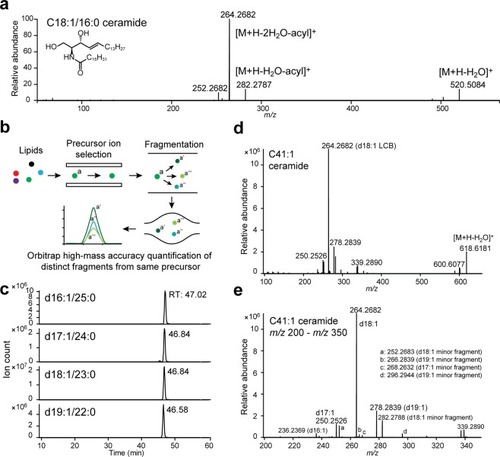
A Parallel Reaction Monitoring-based approach for ceramide quantification. (a) MS2 of d18:1/16:0 ceramide standard in positive ionisation mode. Fragment structures are based on published assignments43,44. (b) Schematic of Parallel Reaction Monitoring. (c) Example PRM chromatograms for C41:1 ceramide from 48 hpf zebrafish embryos, demonstrating presence of multiple LCB-acyl chain isomers with the same m/z. RT: retention time. (d) MS2 spectrum of C41:1 ceramide from 48 hpf zebrafish embryos, the four major LCB fragments are labelled. (d,e) m/z 200-m/z 350, the four major LCB fragments and additional LCB-derived minor fragments are labelled. Unlabelled minor fragments: m/z 224.2371 (d16:1 LCB), m/z 238.2525 (d17:1 LCB).

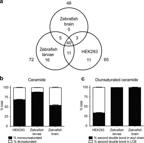
Ceramide composition of zebrafish brain, larvae and HEK239 cells. (a) Venn diagram illustrating total number of quantified ceramides and overlap among different sample types. (b) Percentage of monounsaturated and diunsaturated ceramides in HEK293 cells, zebrafish larvae and brain. (c) Percentage of HEK293, zebrafish larva and brain diunsaturated ceramides with the second degree of unsaturation in either the acyl chain or the LCB. SEM, n = 4 per group. Representative data from three independent experiments are shown.

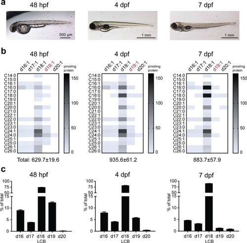
Ceramide regulation during zebrafish embryogenesis. (
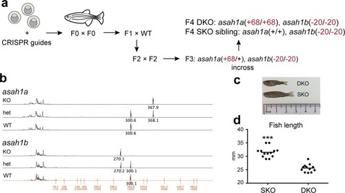
a) Zebrafish at 48 hpf, 4 dpf and 7 dpf. (b) Heatmap of ceramide content in 48 hpf, 4 dpf and 7 dpf larvae, demonstrating time-dependent reduction in the majority of d19:1 (highlighted in red) ceramides (see Table S3 for quantifications). (c) LCB distribution of all detected ceramides from 48 hpf, 4 dpf and 7 dpf larvae. n = 4 per group. Representative data from two independent experiments are shown. Additional verification of LCB distributions in 48 hpf embryos (n = 5 total) was performed via untargeted lipidomics (Supplemental Methods).

A zebrafish model of Farber disease. (a) Schematic of asah1a/b−/− zebrafish generation and propagation. (b) DNA fragment analysis demonstrating presence of altered alleles. The 30-bp difference in the asah1b spectra (rather than the noted 20-bp deletion) is due to presence of different polymorphisms across the introns of the WT and KO alleles (Fig. S2b, see Fig. S3 for sequencing of full cDNA). (c) asah1a/b−/− (DKO) and asah1b−/− (SKO) zebrafish at 3.5 months. (d) Length of DKO and SKO zebrafish at 3.5–4 months. Zebrafish length measurements: Student t-test, SEM, combined data from n = 13 SKO and n = 12 DKO fish, fish within each data set (n = 4–5 SKO, n = 4 DKO) are age-matched. ***p < 0.001.

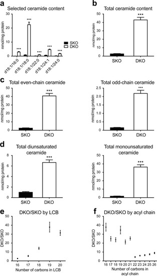
Altered ceramide distribution in Farber disease zebrafish. (a) Quantification for selected ceramides from 3.75-month DKO and SKO zebrafish brains (see Table S4 for quantification of additional ceramide species). (b) Total ceramide content in 3.75-month DKO and SKO zebrafish brains. (c) Total ceramide content by even and odd carbon number. Even: even number of carbons in both acyl chain and LCB; odd: odd number of carbons in acyl chain and/or LCB. (d) Total ceramide content by degrees of unsaturation. Monounsaturated ceramide: one double bond in LCB; diunsaturated ceramide: one additional double bond relative to monounsaturated species. (e) DKO/SKO fold change by LCB length. (f) DKO/SKO fold change by acyl chain length. Student t-test, SEM, n = 4 per group. Representative data from two independent experiments are shown ***p < 0.001.

ZFIN is incorporating published figure images and captions as part of an ongoing project. Figures from some publications have not yet been curated, or are not available for display because of copyright restrictions.

PHENOTYPE:
Fish:
Observed In:
Stage: Adult

Unillustrated author statements

Acknowledgments
This image is the copyrighted work of the attributed author or publisher, and ZFIN has permission only to display this image to its users. Additional permissions should be obtained from the applicable author or publisher of the image. Full text @ Sci. Rep.