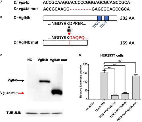
The establishment of a zebrafish vgll4b knockout line. (A) Schematic representation of Cas9 target site in the first exon of zebrafish vgll4b. Dr: Danio rerio. The deleted nucleotides in the mutant gene are marked by hyphens. (B) Schematic representation of wild type (282 amino acids) and mutant Vgll4b proteins (169 amino acids). The site where the frameshift was introduced is marked by triangles. (C) Western blot analysis of HA-tagged wild type and mutant Vgll4b proteins. NC: non-specific control. (D) Luciferase analysis of HEK293T cells transfected with TEAD1, YAP, VGLL4, and wild type or mutant Vgll4b expressing plasmids. Luciferase activity was normalized to empty vector pcDNA3.1 which was set to 1.0. Error bars represent ± SD of at least three replicates. p values are denoted by asterisks. ∗∗∗P < 0.001 (ANOVA test).

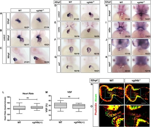
Deficiency of zebrafish vgll4b leads to impaired valvulogenesis. (A–D) WISH analysis of cmcl2 expression in zebrafish embryos at 22, 36, 48, and 96 hpf, a defective S shaped heart looping was clearly observed from 36 hpf. White dashed lines indicate the heart morphology outlined by cmlc2 expression. Lateral view, (C). (E–K) WISH analysis of vmhc, amhc, anf at 48 hpf, as well as bmp4, notch1b, klf2a, spp1 at 52 hpf. (L,M) Optical Heartbeat analysis of cardiac function. Comparison of the heart rate at 48 hpf (L) and ventricular shortening fraction at 52∼54 hfp (M). (N,O) Representative single confocal z-section images showing the endocardial cushion cells expressing Alcam (asterisks) of wild type sibling and vgll4b–/– embryos at 52 hpf. Scale bar: 10 μm.

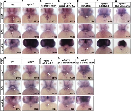
Tead is not involved in the impaired valvulogenesis in vgll4b-deficient embryos. (A–D)Tead1a and tead3b DN mRNA rescue assays in vgll4b–/– embryos. Bmp4, klf2a, and notch1b probes were used in WISH. (E–G)Vgll4b–/– embryos were treated with verteporfin (30 μM) from 32 hpf, but no rescue effect was observed. (H–L)Vgll4b mutants mRNA rescue assays in vgll4b–/– embryos.

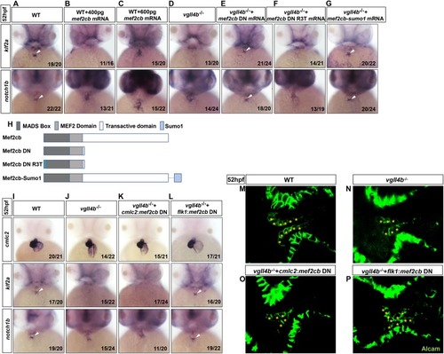
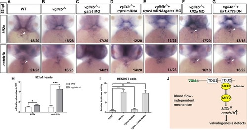
Aberrant activation of Mef2c due to the disruption of Vgll4b-Mef2c complex, accounts for the valvulogenesis defects in vgll4b mutants. (A–C)Mef2cb mRNA was injected into one-cell stage wild type embryos. (D–G)Mef2cb DN, mef2cb DN R3T, and mef2cb-sumo1 mRNA rescue assays in vgll4b–/– embryos. (H) Schematic representation of variant forms of Mef2cb, including WT, DN, R3T, and Sumo1 fusion mutants. (I–L)Cmlc2:mef2cb DN or flk1:mef2cb DN plasmid rescue assays in vgll4b–/– embryos. (M–P) Representative images show Alcam staining of sibling and vgll4b–/– embryos rescued with cmlc2:mef2cb DN or flk1:mef2cb DN plasmid at 52 hpf. The endocardial cushion cells expressing Alcam were indicated by asterisks. Scale bar: 10 μm.

The failure of klf2a expression in the AVC is blood flow-independent, and klf2a is aberrantly activated by Mef2c in the endocardium, which ultimately impedes valvulogenesis. (A–C)Vgll4b–/– embryos were treated with a high dose of gata1 MO, but no rescue effect was observed. (D,E)Trpv4 mRNA was injected alone or in combined with gata1 MO into vgll4b–/– embryos. (F,G)Klf2a MO and flk1:klf2a DN rescue assays in vgll4b–/– embryos. (H) The hearts were harvested respectively from 35 wild type and 35 vgll4b–/– embryos at 52∼54 hfp. Q-PCR analysis revealed that the expression level of klf2a and notch1b in vgll4b–/– hearts was elevated (0.34-fold and 3.2-fold inductions compared to controls). Error bars represent ± SD of at least three replicates. p values are denoted by asterisks; P < 0.1, ∗∗∗P < 0.001 (Student’s t test). (I) Dual luciferase vectors each with a fragment of the zebrafish klf2a promoter (–1.5 kb) were co-transfected into HEK293T cells with empty vector pCS2+, a mef2cb expressing vector, mef2cb combined with wild type vgll4b or vgll4b ΔTDU2 expressing vector. Luciferase activity was detected and normalized to empty vector pCS2+ which was set to 1.0. Error bars represent ± SD of at least three replicates. p values are denoted by asterisks; ∗∗∗P < 0.001 (ANOVA test). (J) Schematic depiction of the aberrant Vgll4b-Mef2c regulation in valvulogenesis in vgll4b–/– zebrafish.

Acknowledgments
This image is the copyrighted work of the attributed author or publisher, and ZFIN has permission only to display this image to its users. Additional permissions should be obtained from the applicable author or publisher of the image. Full text @ Front Cell Dev Biol