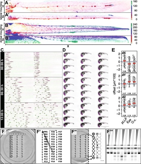
Representative Results.
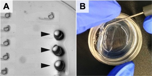
 A Maximum intensity projection (MIP) of a 50 hpf embryo mounted on its side (XY) and corresponding MIP of the orthogonal view (XZ) of the same embryo showing how flat the embryos are mounted in the plate (A’). B MIP of a 40 hpf embryo mounted dorsally (XY) and corresponding MIP of the orthogonal view (XZ) of the same embryo (B′). Depth colour encoding is indicated by colour scales on the right. c Multi-position (36), multi-channel (2) time-lapse recording (13 h duration; 15 min. Interval, see also Additional file 1: Movie S1). D Multichannel (2) Extended Depth of Focus (EDF) projections from widefield Z-stacks (recorded with 20x Objective). Scale Bar = 1 mm (E) Multi-point coordinates in X, Y and Z (recorded with 40x Objective). The offset describes the distance of each point from the mean of all points in X, Y and Z (See methods). Panel 1–3 (top to bottom) show dimensions X, Y and Z in comparison for the pLLP, the eye and the otic vesicle. The red line indicates the median, the blue line indicates zero offset, error bars indicate mean ± standard deviation. Numeric values indicate the variance. F-F″’ Systematic retrieval for genotyping. F Mounted embryos in a 2-D coordinate system of rows (A-M) and columns (1–3). F′ Imaging Sequence in a snake by column fashion. In a time-lapse setting, it starts at point 1 (P01) again to initiate the next timepoint. F″ After imaging, the embryos are retrieved in the same sequence as they were imaged (snake by column, left panel). F”’ Each genotyping result on the electrophoresis gel is easily correlated to one imaging dataset with defined X-Y coordinates

Stamping procedure. a Clean stamp surface b Removal of the 3D-stamp from the polymerized agarose cast using a preparation needle c Ready-to-use mounting cast

Mounting cast under the stereo microscope (a) without LMPA and (b, c) with LMPA with different mirror and contrast adjustment in B (μ-wells not visible) and C (μ-wells visible) (1). inner well and mounting area (2) single μ-well (3) outer well. See also movie S2

Mounted and tail-aligned embryos (9 out of 44). Stripe lines indicate horizontal tail alignment

Imaging setup. a Stage positioned on embryo at position 1 (P01 – upper left corner) b ROI definition and focus refinement in brightfield. c Navigation through the μ-wells by a custom defined well plate. Magenta well is current. Black wells are used to calibrate the well plate

Embryo retrieval. a Arrowheads point to incisions indicating locations where the pipette was introduced into the agarose to retrieve the embryos b Removal of the agarose bed with a preparation needle

Acknowledgments
This image is the copyrighted work of the attributed author or publisher, and ZFIN has permission only to display this image to its users. Additional permissions should be obtained from the applicable author or publisher of the image. Full text @ BMC Biotechnol.