FIGURE SUMMARY
Title

Inhibition of in-stent restenosis after graphene oxide double-layer drug coating with good biocompatibility

Authors
Ge, S., Xi, Y., Du, R., Ren, Y., Xu, Z., Tan, Y., Wang, Y., Yin, T., Wang, G.
Source
Full text @ Regen Biomater

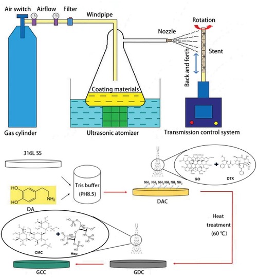
The coating processing and the CS-III pro-ultrasonic spray system [15]. DAC, dopamine coating; GDC, DAC+GO/DTX; GCC, GDC+CMC/Hep

GCC coating has minimum effects on surface roughness but significantly decreases water contact angle. (A) The surface roughness after various coatings. 316L SS stents were coated with DAC, GDC or GCC and then imaged by atomic force microscopy. (B) Different surfaces were imaged by SEM. (C, D) GCC coating significantly decreases the water contact angle. **P < 0.01

GCC Coating exhibits superior blood compatibility. (A, B) GCC coating inhibits the adhesion and activation of platelets. Platelets were plated on 316L SS surfaces with DAC, GDC or GCC for 1 h. The adhered cells were then fixed and imaged by SEM. Rectangular indicates cell spreading and aggregation, while red arrow represents pseudopods. The number of adhered platelets was quantified in (B). the level of platelet activation on the GCC surface (represented by the pseudopodia) was much lower than other three groups, while the GDC coating showed the highest platelet activation. (C) GCC coating exhibits low hemolysis rate. *P < 0.05; **P < 0.01

GDC and GCC Significantly inhibit the proliferation of VSMCs. VSMCs were plated on 316L SS coated with DAC, GDC or GCC. The images were taken at Days 1, 3 and 5, respectively (A). cell proliferation was measured by SEM at various time points accordingly (B). n = 3 independent experiments. **P < 0.01

Various coatings exhibit no detectable toxicity in the embryonic development of zebrafish. Zebrafish embryos were co-cultured with 316L SS, DAC, GDC and GCC stents for 24, 48 and 72 h, respectively. The morphology (A), survival rate (B), hatching rate (C) and heart rate (D) of these embryos were measured at various time points. No significant difference was found among different groups, n = 3

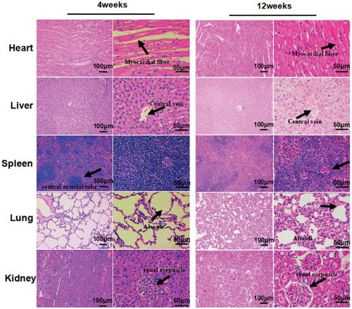
The GCC Stents have no significance effects on the main organs of rabbits at 4 and 12 weeks after implantation. The black arrows represent myocardial fiber, Central vein, Central arterial tube, alveoli and renal corpuscle

The Endothelialization and restenosis of GCC stents. (A) Complete neointima is generated at 4 weeks after implanting 316L SS and GCC stents. Blood components (yellow arrow) adhered and fibrous tissue-like materials (red arrow) appeared on the surface of 316L SS stents. The endothelial cells in GCC exhibited cobblestone-like (purple coils) re-arrangement following the direction of blood flow (purple arrow) at 4 and 12 weeks after stent implantation, respectively. (B) GCC inhibits intimal hyperplasia and the generation of neointima (S, stent; N, neointima; L, lumen; M, media). (C) The cells in intima and media are disordered and elastic plate is not clear or lacking at the distal and proximal sites after implanting 316L SS but not GCC stents

Acknowledgments
This image is the copyrighted work of the attributed author or publisher, and ZFIN has permission only to display this image to its users. Additional permissions should be obtained from the applicable author or publisher of the image. Full text @ Regen Biomater