FIGURE SUMMARY
Title

Inhibited Lipophagy Suppresses Lipid Metabolism in Zebrafish Liver Cells

Authors
Wang, J., Han, S.L., Lu, D.L., Li, L.Y., Limbu, S.M., Li, D.L., Zhang, M.L., Du, Z.Y.
Source
Full text @ Front. Physiol.

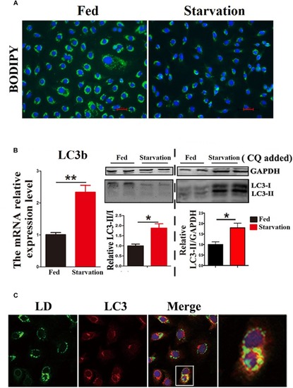
Starvation induces lipophagy in ZFL cell line. (A) Lipid droplets (LDs) stained with BODIPY 493/503 (green) in ZFL cell line in starvation condition. (B) The mRNA expression, immunoblot and densitometric analysis of LC3 in the samples in ZFL cell line. (P < 0.05, ∗∗P < 0.01, n = 6). Error bars, SEM. (C) Co-localization of BODIPY 493/503 (green) with LC3 (red) in ZFL cell line in starvation condition. Nuclei are highlighted with 4, 6-diamidino-2-phenylindole (DAPI).

Electron micrographs of ZFL cells. (A,B) The structure of an autophagic vesicole (AV). (C–H) The structure of an autophagosome containing a lipid droplet (AP). N, nucleus; ER, endoplasmic reticulum; Mit, mitochondria.

Inhibited lipophagy causes accumulation of lipid droplets in ZFL cell line. (A,B) Lipid droplets (LDs) stained with BODIPY 493/503 and TG contents in the ZFL cells. (C) Immunoblot and densitometric analysis of LC3 and p62 in the ZFL cells. (P < 0.05, n = 6). Error bars, SEM. (D) Co-localization of BODIPY 493/503 (green) with LC3 (red) in treated samples with CQ.

Acknowledgments
This image is the copyrighted work of the attributed author or publisher, and ZFIN has permission only to display this image to its users. Additional permissions should be obtained from the applicable author or publisher of the image. Full text @ Front. Physiol.