
- Title
-
Bar, stripe and spot development in sand-dwelling cichlids from Lake Malawi
- Authors
- Hendrick, L.A., Carter, G.A., Hilbrands, E.H., Heubel, B.P., Schilling, T.F., Le Pabic, P.
- Source
- Full text @ EvoDevo

Adult pigmentation in C. azureus and D. compressiceps. a Vertical bars b1–9, interbars i1–8 and spots S1–3 in C. azureus. b, c Higher magnification images of bars/interbars and spots in C. azureus before (b) and after (c) adrenaline treatment. d, e Melanophores (black), xanthophores (orange) and iridophores (silver) in bars and interbars of C. azureus before (d) and after (e) adrenaline treatment. f, g Melanophores, xanthophores and iridophores of spot in C. azureus before (f) and after (g) adrenaline treatment. h Melanophore and xanthophore average densities in bars, interbars and spots (n = 3) in C. azureus. Error bars represent standard deviations. i Melanophore darkness in spot and bar expressed as integrated density. j Stripes and interstripes in D. compressiceps adult. k, l Higher magnification images of stripes and interstripes in D. compressiceps flank before (k) and after (l) adrenaline treatment. m–o Melanophores, xanthophores and iridophores of stripes SDL and SML and interstripe X1DL before (k, m) and after (l, n) adrenaline treatment. o Lack of xanthophores in interstripe X1DL visualized under fluorescent light (488 nm). p Melanophore and xanthophore average densities in SDL, SML and SVL (n = 3) in D. compressiceps. Error bars represent standard deviations. a, j Scale = 1 cm. b, d, k, l Scale = 1 mm. d–g and m–o Scale = 250 µm |

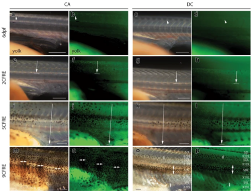
Larval development of pigmentation in C. azureus and D. compressiceps. a–c, e–g Pre-metamorphic development in C. azureus and D. compressiceps larvae. a, e Melanophores on the yolk (arrows) and melanization of the pigmented epithelium of the eye (arrowheads) in C. azureus and D. compressicepsat 4dpf. c, g Melanophores over the dorsal head in C. azureus and D. compressiceps (insets) and iridophores in the eye in C. azureus and D. compressiceps (arrowheads) at 6dpf. d, h–x Development of metamorphic pigmentation in C. azureus and D. compressiceps. d, h Myotome morphology at the onset of overt metamorphosis in C. azureus and D. compressiceps at 7dpf. i, m Melanophores over the dorsal neural tube in C. azureus and D. compressiceps (insets). j, n Skin melanophores at the base of the dorsal fin (arrowheads) and in C. azureus trunk and tail and D. compressiceps tail (insets). k, l, o, p Additional skin melanophores in C. azureus an D. compressiceps. l, p Iridophores in C. azureus and D. compressiceps in posterior caudal fin peduncle (arrowheads). l Dorsal melanophore patches D1–3 in C. azureus. q Dorsal patch D4 on caudal peduncle. r Dorsal patch D1 into D1a and D1b, dorsal patch D2 into D2a, D2b; lateral patches P4 and P5; and iridophores between melanophore patches (inset) in C. azureus. s Lateral melanophore patch P6 in C. azureus. u Melanophores at anterior portion of presumptive midlateral stripe SML (arrow), at presumptive dorsolateral stripe SDL (arrowhead) and presumptive ventrolateral stripe SVL (inset) in D. compressiceps. v Iridophores in presumptive ventrolateral interstripe 1 X1VL (inset). w Iridophores in presumptive dorsolateral interstripe 1 X1DL (arrowhead) in D. compressiceps. x Complete dark stripe pattern at late larval stage in D. compressiceps. Scale = 1 mm |

Morphological changes in median fins and myotomes during metamorphosis in D. compressiceps. aBulges (arrows) in embryonic median fin prefigure the posterior ends of the dorsal and anal fin, respectively. “V” shape of myotomes indicated in white. b Unsegmented caudal fin ray elements. cDorsal- and anal-fin ray condensations indicated by short arrows. Chevron shape of myotomes indicated by long arrows. d 2 caudal fin ray elements separated by one joint (white arrow). e Fin ray elements lengthen in dorsal fin and anal fin (short arrows). Melanophores appear on dorsal neural tube (long arrow). f 3 caudal fin ray elements separated by two joints (white arrows). g Dorsal and anal fins separated from caudal fin (short arrows). Melanophores appear in flank skin. h 4 caudal fin ray elements separated by three joints (white arrows). i Progressive disappearance of larval fin tissue from dorsal and ventral caudal fin peduncle. j 5 caudal fin ray elements separated by four joints (white arrows). The timing of metamorphosis is the same in C. azureus as in D. compressiceps. Scale = 500 µm |
Xanthophore distributions in C. azureus and D. compressiceps larvae. a–d Xanthophores are restricted to the dorsal midline in C. azureus and D. compressiceps at 6dpf (arrows) as visualized with white light (a, c) or 488 nm fluorescent light (b, d). e–h Xanthophores extend from dorsal midline to horizontal myoseptum (arrow) in C. azureus and D. compressiceps as visualized with white light (e, g) or 488 nm fluorescent light (f, h). i–l Xanthophores extend from dorsal to ventral midlines (arrow) in C. azureusand D. compressiceps as visualized with white light (i, l) or 488 nm fluorescent light (j, l). m–nXanthophore density is greater under melanophore patches (double-headed arrows) in C. azureus at 17dpf/9CFRE. o–p Xanthophores are largely restricted to dark stripes (double-headed arrows) in D. compressiceps at 17dpf/9CFRE. Scale = 500 µm |

Melanophore and iridophore patterns in C. azureus and D. compressiceps larvae. a Melanophores in the flank in C. azureus—patches P1–3 and base of dorsal fin (arrow). b Melanophores along tail lateral midline and base of dorsal fin (arrow) in D. compressiceps. c Melanophore additions to and around P1–3 in C. azureus; Resolution of dorsal midline melanophore band into D1, D2 and D3. Appearance of iridophore patch on dorsal caudal fin peduncle (arrow). d Melanophore additions to tail-portion of dark stripe S0 in D. compressiceps. Iridophore patch on dorsal caudal fin peduncle (arrow). eResolution of dorsal melanophore patches D1 into D1a and D1b, and of D2 into D2a, D2b; appearance of D4 on dorsal caudal fin peduncle. Appearance of lateral patches P4 and P5. Iridophore patches between dorsal and lateral patches (arrowheads). f Melanophores along trunk portion of SML in D. compressiceps (black bracket). Melanophores at presumptive SDL and SVL (arrowheads). Iridophores dorsal and ventral to horizontal myoseptum (white brackets). g New melanophores around populated areas. Appearance of lateral melanophore patch P6 in C. azureus. h Iridophores become abundant at interstripes X1DL and X1VL in D. compressiceps. Scale = 1 mm |

Timelapse recording of bar melanophore development in C. azureus. a–i Melanophores of presumptive lateral patch P4 (left) and a posteriorly appearing patch over 6 days of development. Melanophores present at 10dpf (a) and 11dpf (b) are identified by the same letters at all timepoints. c New melanophores are circled in red at this stage (12dpf early), and by red dots at subsequent stages. White arrowheads indicate daughter cells produced by melanophore mitosis. d New melanophores are circled in dark blue at this stage (12dpf late), and by dark blue dots at subsequent stages. e New melanophores are circled in light blue at this stage (13dpf early), and by light blue dots at subsequent stages. f New melanophores are circled in yellow at this stage (13dpf late), and by yellow dots at subsequent stages. Black arrowheads indicate daughter cells produced by melanophore mitosis. g New melanophores are circled in purple at this stage (14dpf), and by purple dots at subsequent stages. Black arrowheads indicate daughter cells produced by melanophore mitosis. h New melanophores are circled in green at this stage (15dpf), and by green dots at subsequent stages. i The relative positions of all melanophores examined remained largely unchanged from 10 to 16dpf. Scale = 250 µm |

Expression of neural crest (NC) cell- and pigment progenitor markers during embryonic development and later in metamorphosis in C. azureus. a–m Whole mount in situ hybridizations (ISH), anterior to the left, dorsal view (a–l) and lateral view (m). a, b foxd3 and sox10 expression in presumptive hindbrain, mandibular- and post-otic NC streams at 36hpf. c–f, h, i foxd3 and sox10 expression in trunk NC cells migrating through the lateral- and medial routes at 42 and 48hpf. Foxd3 is also expressed in somites. g ednrb1a expression in trunk NC cells migrating through the medial route at 48hpf. h–iHigher magnifications of foxd3 and sox10 expression in trunk NC cells migrating through the lateral- and medial routes at 48hpf. j–k foxd3 and sox10 expression in trunk NC cells migrating through the lateral- and medial routes at 54hpf. Foxd3 expression is also detected in somites. l ednrb1a expression in trunk NC cells migrating through the medial route at 54hpf. m Higher magnification of ednrb1aexpression in trunk NC cells migrating through the lateral route at 48hpf (somites 1–9 numbered). n–oISH on cryosections of 11dpf/5CFRE in C. azureus larvae. Ednrb1a and mitfa expression is detected at high levels in segmented structures located dorsal to the neural tube (#), and at lower levels along myotome boundaries (arrowheads) and in the skin. e eye, hb hindbrain, lr lateral NC route, mbmyotome boundary, md mandibular NC stream, mr medial NC route, my myotome, ov otic vesicle, post-otic post-otic NC stream, s somite. Scale = 250 µm |