
- Title
-
Spatio-temporal characterization of dynamic epithelial filopodia during zebrafish epiboly
- Authors
- Rutherford, N.E., Wong, A.H., Bruce, A.E.E.
- Source
- Full text @ Dev. Dyn.
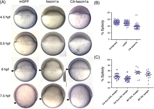
Expression of constitutively active fascin1a delays epiboly initiation. A, Lateral views of live embryos during epiboly, times and injected constructs as indicated. Black arrows indicate the margin. Data generated from six experiments with approximately 40 embryos per group. B, Percent epiboly at 6 hpf, data from five experiments. One‐way ANOVA between uninjected, mGFP, and CA fascin1a: P < .0001. Error bars are SD. C, EVL and deep cells were both delayed in CA‐fascin1a‐injected embryos. Fascin1a N = 20, wild type N = 15. Error bars are SEM. t‐Tests were performed between CA fascin1a EVL margin with WT fascin1a EVL margin, and CA‐fascin1a DEL margin with WT‐fascin1a DEL margin: P = .0021 and .0016, respectively |