
- Title
-
CNBP controls transcription by unfolding DNA G-quadruplex structures
- Authors
- David, A.P., Pipier, A., Pascutti, F., Binolfi, A., J Weiner, A.M., Challier, E., Heckel, S., Calsou, P., Gomez, D., Calcaterra, N.B., Armas, P.
- Source
- Full text @ Nucleic Acids Res.
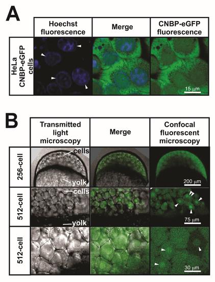
Subcellular localization of CNBP in HeLa cells and zebrafish embryos. A. Localization of CNBP-eGFP in HeLa CNBP-eGFP live cells. Images generated by Hoechst staining (left) and GFP fluorescence (right) using confocal fluorescent microscopy. Merged images are shown in the middle. White arrowheads point fluorescent nuclei. B. Localization of zCnbp-eGFP in live zebrafish embryos overexpressing zCnbp-eGFP staged prior (256-cell) and after (512-cell) mid-blastula transition (MBT). Images generated by transmitted light (left) and confocal fluorescent (right) microscopy are shown. Merged images are shown in the middle. White arrowheads point fluorescent nuclei. Upper and middle panels show lateral views of embryos (animal pole to the top) while lower panels show a detail with cells from the animal pole. Materials and Methods: (A) Hoechst staining was performed on live HeLa CNBP-eGFP cells as detailed elsewhere (6). Confocal microscopy was performed using a ZEISS LSM 710 confocal laser scanning microscope equipped with a 40X/1.3 oil immersion objective. (B) live zebrafish embryos overexpressing zCnbp-eGFP were documented using a Nikon TE2000-E confocal microscope with an eclipse C1 head. |