FIGURE SUMMARY
Title

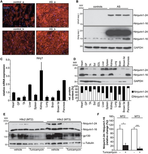
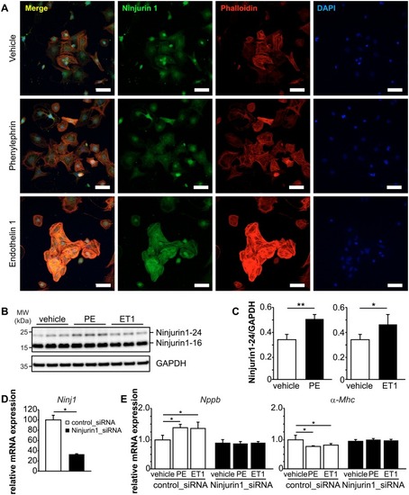
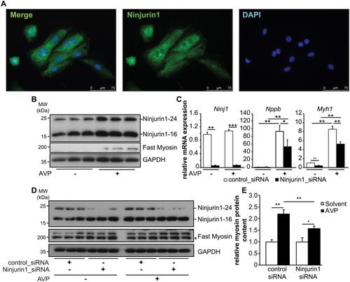
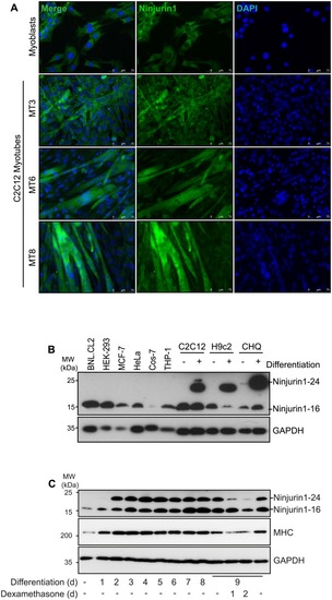
Ninjurin1 regulates striated muscle growth and differentiation

Authors
Kny, M., Csályi, K.D., Klaeske, K., Busch, K., Meyer, A.M., Merks, A.M., Darm, K., Dworatzek, E., Fliegner, D., Baczko, I., Regitz-Zagrosek, V., Butter, C., Luft, F.C., Panáková, D., Fielitz, J.
Source
Full text @ PLoS One

Ninjurin1 is increased in hypertrophic left ventricle of patients with severe aortic stenosis.

(A) Immunofluorescent staining of cryosections from biopsy specimen of two AS (AS_a, AS_b) and two donor (control_a, control_b) hearts with anti-Ninjurin1 as primary antibody and Alexa Fluor 555 conjugated secondary antibody. Nuclei were stained with DAPI (blue). MW, molecular weight; kDa, kilo Dalton. Scale bar, 75 μm. (B) Western blots of proteins isolated from left ventricular tissue of patients with severe aortic stenosis (AS) undergoing elective aortic valve replacement surgery (n = 3) and donor hearts (n = 3) using anti-Ninjurin1 antibody. GAPDH was used as loading control. The Ninjurin1 isoforms at 16kDa (Ninjurin1-16) and 24kDa (Ninjurin1-24) are differentially expressed therefore the membranes underwent short and long exposure, indicated by short exp. and long exp., respectively. (C) Quantitative RT-PCR analysis of Ninjurin1 (Ninj1) in different mouse tissues obtained from untreated C57BL/6J mice as indicated (n = 3). 18S ribosomal RNA expression was used as reference. Data are presented as mean ± SD. (D) Representative immunoblot of proteins isolated from different mouse tissues using anti-Ninjurin1 antibody. GAPDH was used as loading control. The Ninjurin1 isoforms at 16kDa (Ninjurin1-16) and 24kDa (Ninjurin1-24) are indicated. Graph: Densitometric analysis of (B). The total Ninjurin1 content (Ninjurin1-16 plus Ninjurin1-24) per tissue was set as 100% and the relative amount of Ninjurin1-16 and Ninjurin1-24, respectively, was related to that. GP indicates gastrocnemius / plantaris; TA, tibialis anterior; Sol, soleus; EDL, extensor digitorum longus. (E) Western blots of proteins isolated from 2 days (MT2) or 3 days (MT3) differentiated H9c2 cells, treated with 1μg/ml Tunicamycin or vehicle (1 mg/ml dimethylsulfoxide) for 48 h, as indicated. Detection of Ninjurin1 and glycoprotein 130 (gp130) using specific antibodies. Tubulin was used as loading control. (F) Densitometric analysis of (E); data are presented as mean ± SD. * P = 0.05; *** P < 0.001. A two-tailed, unpaired Student’s t-test was used to calculate the P values.

Knockdown of Ninjurin1 by siRNA impairs cardiomyocyte hypertrophy <italic>in vitro</italic>.

(A) Immunofluorescent staining of NRVM treated with vehicle, phenylephrine (100 μM) and endothelin 1 (50 nM) with anti-Ninjurin1 as primary antibody and Alexa Fluor 488 conjugated secondary antibody (green). Nuclei were stained with DAPI (blue). Scale bar, 75 μm. (B) Immunoblots (left) and densitometric analysis (right) of proteins isolated from vehicle, phenylephrine (100 μM) and endothelin 1 (50 nM) treated NRVM using anti-Ninjurin1 antibody. GAPDH was used as loading control. The 16kDa (Ninjurin1-16) and 24kDa (Ninjurin1-24) Ninjurin1 isoforms are indicated. (C) Densitometric analysis of the Ninjurin1-24 signal from (B). (D, E) NRVM were transfected with control siRNA (control_siRNA) (n = 3) or siRNA targeting Ninjurin1 (Ninjurin1_siRNA) (n = 3). (D) Quantitative RT-PCR analysis of Ninjurin1 (Ninj1). GAPDH expression was used as reference. Data are presented as mean ± SD. * P < 0.05. (E) Quantitative RT-PCR analysis of Nppb and alpha-myosin heavy chain 1 (α-Mhc). GAPDH expression was used as reference. Data are presented as mean ± SD. * P < 0.05, ** P < 0.01, *** P < 0.001.

Knockdown of Ninjurin1 by siRNA leads to impaired cardiomyocyte hypertrophy <italic>in vitro</italic>.

(A) Immunofluorescent staining of H9c2 myocytes with anti-Ninjurin1 as primary antibody and Alexa Fluor 488 conjugated secondary antibody (green). Nuclei were stained with DAPI (blue). Scale bar, 75 μm. (B) Hypertrophy of 7 days differentiated H9c2 myotubes was induced by arginine-vasopressin (AVP) treatment for 24 hours. Non-treated cells were used as control. Immunoblots of proteins isolated from H9c2 myotubes (as indicated) using anti-Ninjurin1 and anti-fast myosin antibody. GAPDH was used as loading control. The 16kDa (Ninjurin1-16) and 24kDa (Ninjurin1-24) Ninjurin1 isoforms are indicated. (C-E) 7 days differentiated H9c2 myotubes were transfected with control siRNA (control_siRNA) (n = 6) or siRNA targeting Ninjurin1 (Ninjurin1_siRNA) (n = 6). 48 hours after transfection hypertrophy was induced by AVP treatment for 24 hours (n = 3). Non-treated cells were used as control (n = 3). (C) Quantitative RT-PCR analysis of Ninjurin1 (Ninj1), Nppb and Myosin heavy chain 1 (Myh1). GAPDH expression was used as reference. Data are presented as mean ± SD. * P < 0.05, ** P < 0.01, *** P < 0.001(D) Immunoblots of proteins isolated from H9c2 myotubes (as indicated) using anti-Ninjurin1 and anti-fast myosin antibody. GAPDH was used as loading control. The 16kDa (Ninjurin1-16) and 24kDa (Ninjurin1-24) Ninjurin1 isoforms are indicated. (E) Densitometric analysis of fast myosin protein content from (D). Data are presented as mean ± SD. * P < 0.05, ** P < 0.01. MW, molecular weight; kDa, kilo Dalton. A two-tailed, unpaired Student’s t-test was used to calculate the P values.

Ninjurin1 increases during myogenic differentiation in skeletal myocytes.

(A) Immunofluorescent staining of C2C12 myoblasts, and for 3, 6 and 8 days differentiated myotubes, as indicated (MT3, MT6, MT8), with anti-Ninjurin1 as primary antibody and Alexa Fluor 488 conjugated secondary antibody (green). Nuclei were stained with DAPI (blue). Scale bar, 75 μm. (B) Western blots of proteins isolated from different non-myocyte (BNLCL2, HEK-293, MCF-7, HeLa, Cos-7, THP-1) and myocyte (C2C12, H9c2, CHQ) cell lines (as indicated) using anti-Ninjurin1 antibody. The myocyte cell lines C2C12, H9c2 and CHQ were analyzed as undifferentiated myoblasts (-) and differentiated myotubes (+). GAPDH was used as loading control. The 16kDa (Ninjurin1-16) and 24kDa (Ninjurin1-24) Ninjurin1 variants are indicated. (C) Western blots of proteins isolated from C2C12 myoblasts and for different time points (as indicated) differentiated myotubes using anti-Ninjurin1 antibody. To induce atrophy 9 days differentiated C2C12 myotubes were treated with Dexamethasone (10 μM) for 1 and 2 days, as indicated. GAPDH was used as loading control. The 16kDa (Ninjurin1-16) and 24kDa (Ninjurin1-24) Ninjurin1 isoforms are indicated. MHC indicates myosin heavy chain.

Ninjurin1 deficiency impairs zebrafish heart and skeletal muscle development.

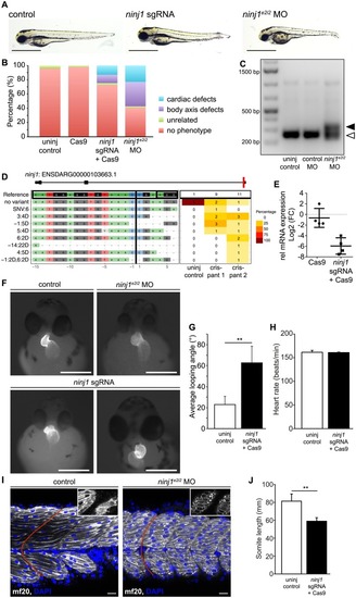
Zebrafish (wild type or transgenic Tg(myl7:EGFP)twu34) embryos were injected with ribonucleoprotein complex of Cas9 and sgRNA targeting exon 1 of ninjurin1 (ninj1 sgRNA) or morpholino targeting exon2/intron2 junction of ninjurin1 (ninj1e2i2 MO) at one cell stage and compared to uninjected control. In all panels, embryos were analysed at 48–50 hours post fertilization (hpf). (A) Representative images of uninjected controls and ninjurin1-deficient embryos showing cardiac and body axis defects. Scale bar, 1 mm. (B) The relative occurrence of the cardiac phenotypes is depicted. (C) qRT-PCR of ninj1 cDNA from uninjected control, control morpholino injected (control MO), and ninje2i2 MO injected embryos at 50 hpf. In control MO injected embryos, the size of the ninj1 amplified fragment is 254 bp (empty arrowhead), whereas it is approximately 350 bp in ninj1 morphants, due to defective splicing of intron 2 (full arrowhead). (D) Panel plot showing allele variations in ninj1 sgRNA-Cas9-injected embryos compared to the WT ninj1 allele (no variant) (ENSDARG000001036663.1). (E) Relative mRNA expression of the ninj1 gene in uninjected, Cas9, and ninj1 sgRNA-Cas9-injected embryos by qRT-PCR and normalized to eef1α1l mRNA expression. Data represent the log2 fold change of ninj1. Cas9 to uninjected control, ns, P = 0.0686, ninj1 sgRNA-Cas9-injected embryos to uninjected controls, **, P = 0.0025. (F) Representative images of Tg(myl7:EGFP)twu34 showing defects in cardiac looping and dextrocardia in ninj1-deficient embryos. Scale bar, 1 mm. (G) Quantification of the looping angle, ninj1 sgRNA-Cas9-injected embryos to uninjected control, **, P = 0.0012. (H) Heart rate remained unchanged in ninj1-deficient embryos; ninj1 sgRNA-Cas9-injected embryos to uninjected control, ns, P = 0.8406. (I) Single confocal plane of whole-mount embryos showing skeletal muscle cells and their nuclei stained with anti-myosin heavy chain antibody (mf20) and DAPI, respectively. Myosin assembles within the sarcomeres in controls, while it forms aggregates in ninj1-deficient embryos (in insets, apical section). Somites do not display the typical chevron shape, red line. Scale bar, 20 μm. (J) Somite length was significantly shorter in ninj1-deficient embryos compared to controls; **, P = 0.0025.

Acknowledgments
This image is the copyrighted work of the attributed author or publisher, and ZFIN has permission only to display this image to its users. Additional permissions should be obtained from the applicable author or publisher of the image. Full text @ PLoS One