
- Title
-
Tracing of Afferent Connections in the Zebrafish Cerebellum Using Recombinant Rabies Virus
- Authors
- Dohaku, R., Yamaguchi, M., Yamamoto, N., Shimizu, T., Osakada, F., Hibi, M.
- Source
- Full text @ Front. Neural Circuits
The
Construct:
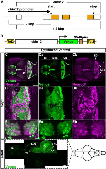
Tg(cbln12:Venus)
|

Transgenic lines for tracing Purkinje cell (PC) afferents. |

Transgenic lines for tracing GC afferents. |

Tracing of the presynaptic neurons of PCs using the RV method. A solution of pseudotyped RV was injected into the left side of the cerebellum of adult |

Tracing of mossy fibers (MFs) using the RV method. The RV solution was injected into the left side of the cerebellum of adult |