
- Title
-
The Dietary Flavonoid, Luteolin, Negatively Affects Neuronal Differentiation
- Authors
- Swaminathan, A., Basu, M., Bekri, A., Drapeau, P., Kundu, T.K.
- Source
- Full text @ Front. Mol. Neurosci.
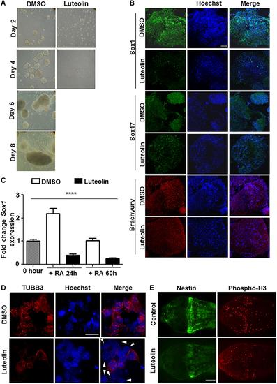
Luteolin treatment perturbs early embryonic stem cell differentiation to the neuronal lineage. (A) Bright-field images of EBs treated with DMSO/luteolin acquired every alternate day show that luteolin treatment strongly prevents EB formation, while DMSO treated control mESCs formed steadily growing EBs. Images of luteolin treated cells beyond day 4 are not shown, since they did not grow any further (N = 2, n = 100 EBs per treatment per time point). (B) Immunofluorescence analysis of germline marker expression in EBs treated with DMSO or luteolin (15 μM) for 24 h from 48 h. The expression of Sox1 (ectodermal marker), Sox17 (endodermal marker), and Brachyury (mesodermal marker) were analyzed. Scale bar: 50 μm (N = 2, n = 3–5 EBs imaged per treatment per marker). (C) qRT-PCR analysis of ESCs treated with retinoic acid (0.5 μM) and DMSO or luteolin (15 μM) for 24 and 60 h to quantify expression of early neuronal marker Sox1. DMSO treated undifferentiated ESCs were taken as control, GAPDH expression was used for normalization. While the expression of both markers increased with time in DMSO and RA treated ESCs, luteolin treatment led to a significant reduction in the expression of both markers. One-way ANOVA performed for the factor of treatment condition: ∗∗∗∗p< 0.0001 (N = 2, n = 3 per treatment). (D) Immunofluorescence analysis of neuronal marker expression in 2-day old EBs grown in the presence of 1 μM RA and differentiated further for 5 days with DMSO or luteolin (15 μM). While β-III tubulin was uniformly expressed in all the DMSO treated control differentiating cells, many luteolin treated cells did not express the neuronal marker (indicated using white arrowheads). Scale bar: 10 μm (N = 2; n ≥ 5 fields per treatment). (E)Immunofluorescence analysis of the expression of Nestin (neuronal marker) and phosphorylated histone H3 (marker of proliferating cells) in 48 h post fertilization zebrafish embryos treated with DMSO or luteolin (100 μM) from 8 hpf. Luteolin treatment led to less cell division in the brain and lesser expression of Nestin itself. Scale bar: 40 μm (N = 2, n = 6–8 zebrafish larval heads per treatment). |