
FIGURE SUMMARY
- Title
-
Zebrafish Embryo Vessel Segmentation Using a Novel Dual ResUNet Model
- Authors
- Zhang, K., Zhang, H., Zhou, H., Crookes, D., Li, L., Shao, Y., Liu, D.
- Source
- Full text @ Comput Intell Neurosci
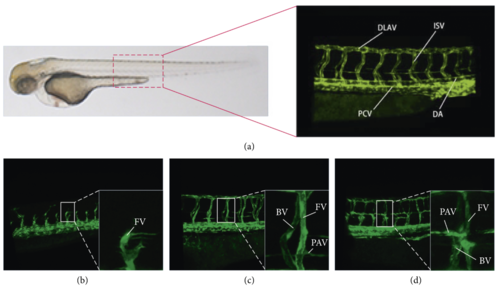
Zebrafish fluorescent vascular 2D view: (a) natural pattern and enlarged view of trunk (DLAV: dorsal; ISV: intersegmental vessel; PCV: posterior cardinal vein; DA: dorsal aorta); (b–d) the zebrafish embryo’s trunk vessels of 35H, 48H, and 72H, respectively (FV: foreground vessel; BV: background vessel; PAV: parachordal vessel). |
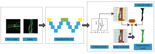
The workflow of the proposed learning methodology. |
Acknowledgments
This image is the copyrighted work of the attributed author or publisher, and
ZFIN has permission only to display this image to its users.
Additional permissions should be obtained from the applicable author or publisher of the image.
Full text @ Comput Intell Neurosci