
- Title
-
Human melanoma brain metastases cell line MUG-Mel1, isolated clones and their detailed characterization
- Authors
- Heitzer, E., Groenewoud, A., Meditz, K., Lohberger, B., Liegl-Atzwanger, B., Prokesch, A., Kashofer, K., Behrens, D., Haybaeck, J., Kolb-Lenz, D., Koefeler, H., Riedl, S., Schaider, H., Fischer, C., Snaar-Jagalska, B.E., de'Jong, D., Szuhai, K., Zweytick, D., Rinner, B.
- Source
- Full text @ Sci. Rep.
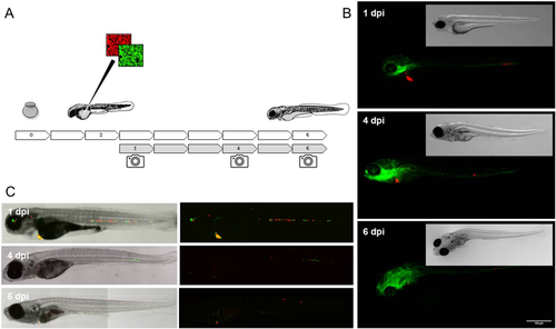
Zebra Fish. Schematic overview of the experimental approach, embryos are collected at day 0 and injected at 2 days post fertilization (dpf) through the embryonic common cardinal vein (Duct of Cuvier), following the embryos are imaged on 1, 4 and 6 days post injection (dpi) to follow the initial dispersal of the cells at 1 dpi, the stage of engraftment around 4 dpi and eventual successful outgrowth of micro metastatic foci at 6 dpi. (A) Fluorescent micrographic images of fli:GFP blood and lymphatic vessel reporter zebrafish engrafted with MUG-Mel1 (red), imaged on day 1, 4 and 6 dpf show dispersal, engraftment and outgrowth of experimental micro metastases. (B) Confocal stich of ABTL zebrafish embryos engrafted with 1:1 mixed clones, C8 (green) and D5 (red), after injection clone C8 clearly does not form any multicellular foci whereas D5 establishes multiple foci 6 days after engraftment. (C) All images represent median phenotypes of injected cohorts, scale bars represent 200 µm.
|