
- Title
-
Global Expression Profiling Identifies a Novel Hyaluronan Synthases 2 Gene in the Pathogenesis of Lower Extremity Varicose Veins
- Authors
- Hsieh, C.S., Tsai, C.T., Chen, Y.H., Chang, S.N., Hwang, J.J., Chuang, E.Y., Wu, I.H.
- Source
- Full text @ J Clin Med
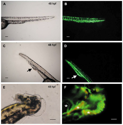
Knockdown of hyaluronan synthases 2 gene (HAS2) in zebrafish embryos leads to defective venous structure with blood flow stasis. Tg(fli1:egfp)xTg(gata1:dsRed) zebrafish with green fluorescence emitting vasculature and red fluorescence emitting red blood cells are used to observe the anatomic change of vasculature (green fluorescence) and blood flow (red fluorescence). (A) Morphology of the tail of a representative wild-type zebrafish (48 hpf) under bright field microscope; (B) Vascular structure of the same zebrafish tail under fluorescence microscope; (C) Morphology of the tail of a representative HAS2-morpholino (MO) injected zebrafish (48 hpf) under bright field microscope. A protruding bulb in the tail is noted (black arrow), mimicking human phenotype of varicose vein; (D) Vascular structure of the same HAS2-MO injected zebrafish tail under fluorescence microscope. A corresponding local dilation of venous structure in the caudal vein plexus (CVP) is found (white arrow); (E) Morphology of a representative HAS2-morpholino (MO) injected zebrafish (48 hpf) with tail malformation under bright field microscope; (F) Vascular structure of the same HAS2-MO injected zebrafish tail under fluorescence microscope. Tangled veins are found in the mal-formatted tail (white stars). Magnification is 100× in (A–D) and 200× in (E,F). Scale bar = 100 μm. Hpf indicates hours post-fertilization. PHENOTYPE:
|
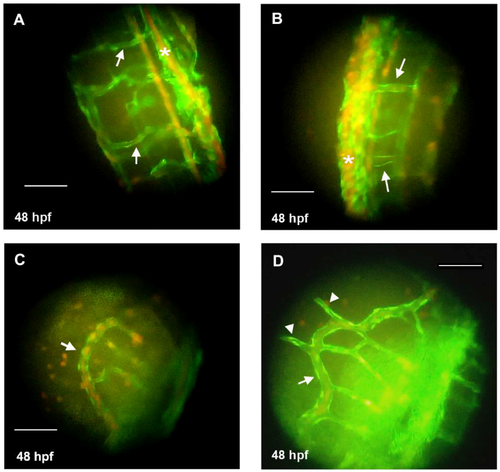
Knockdown of hyaluronan synthases 2 gene (HAS2) in zebrafish embryos leads to dilated venous structure. Tg(fli1:egfp)xTg(gata1:dsRed) zebrafish with green fluorescence emitting vasculature are used to observe the anatomic change of vasculature. (A) Inter-segmental veins (ISVs) in the tail of a representative wild-type zebrafish (48 hpf) under fluorescence microscope (arrows); (B) Dilated ISVs in the tail of a representative HAS2-morpholino (MO) injected zebrafish (48 hpf) under fluorescence microscope (arrows); (C) The sub-intestinal vein (SIV) in the tail of a representative wild-type zebrafish (48 hpf) under fluorescence microscope (arrow); (D) The dilated SIV (arrow) with protruding branches (arrowheads) in the tail of a representative HAS2-morpholino (MO) injected zebrafish (48 hpf) under fluorescence microscope. Hpf indicates hours post-fertilization. Magnification is 400×. Scale bar = 100 μm. PHENOTYPE:
|