
- Title
-
Deletion of Pr72 causes cardiac developmental defects in Zebrafish
- Authors
- Song, G., Han, M., Li, Z., Gan, X., Chen, X., Yang, J., Dong, S., Yan, M., Wan, J., Wang, Y., Huang, Z., Yin, Z., Zheng, F.
- Source
- Full text @ PLoS One
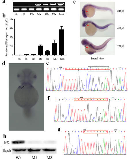
Expression patterns of pr72 and verification of pr72 knockout. (a, b) Time course expression of pr72 in zebrafish embryos at 0, 6, 12, 24, 48, 72 hpf and in adult hearts, identified by RT-PCR (a) and qPCR (b). (c) Expression of pr72 at 24, 48 and 72 hpf, as shown by whole mount in situ hybridization. A lateral view, anterior to the left, the expression in the heart is shown by red arrows. (d) A ventral view of the in situ result of embryos at 72 hpf, anterior to the top, the expression in the heart is indicated by a black arrow. (e, f & g) The sequencing results of TALEN target sites (indicated by the red frame) in PCR products amplified from F2 zebrafish (M1 (f) and M2 (g)) and the wild type (Wt); the restriction enzyme ScaII recognition site was shown using a black box in Wt. (h) The Pr72 Western-blot results of Wt, M1 and M2. |
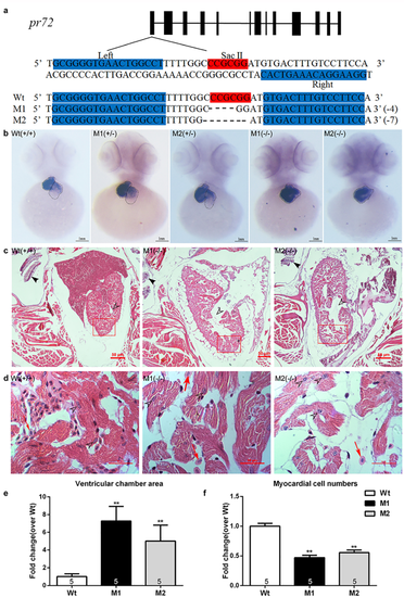
Pr72 KO mediated by TALEN induced cardiac phenotypes in zebrafish. (a) The TALEN target site of exon 1 of the zebrafish pr72 gene. The binding sites used in this study (indicated by “Left” and “Right”) are highlighted in cyan; the SacII site in the spacer is highlighted in red. Representative sequencing results revealed different InDels in the TALEN target site. (b) WISH of cmlc2 expression shows the ventricles and atria in zebrafish embryos, and the ventricles and atria were circled by a dotted line while the ventricle was on the left and the atrium was on the right. The ventricles were enlarged, while the atrial wall was thinning and the atrium was even invisible in the homozygous fish compared to the heterozygous and Wt fish. (c, d) Representative histopathologic sections stained with HE at 40×(c) and 1000×(d), Scale bars 50μM. (c) Representative Hollow arrowheads indicated the ventricular chambers. The bony landmarks pointed by solid arrowheads indicated that the slices were from the same region of the fish body. (d) Higher magnification regions in the red box (1000×). The heart tissues Wt, M1 and M2 were at the same section of the heart. Hollow arrowheads pointed the myocardium nucleus. Red arrows indicated the erythrocytes. (e) Quantification of ventricular chamber areas normalized to ventricular areas, and compared to Wt. (f) Quantification of myocardium cell numbers compared to the control. Myocardium amounts were counted in each of 10 randomly chosen fields per fish at a magnification of 1000× by two observers blinded to the identification of the fish from which the images were obtained. Data are expressed as mean ± SEM. **, p < 0.01 vs controls, one-way ANOVA followed by Dunnett’s posttest. The numbers of zebrafish are indicated in the columns. PHENOTYPE:
|

ZFIN is incorporating published figure images and captions as part of an ongoing project. Figures from some publications have not yet been curated, or are not available for display because of copyright restrictions. PHENOTYPE:
|
|
ZFIN is incorporating published figure images and captions as part of an ongoing project. Figures from some publications have not yet been curated, or are not available for display because of copyright restrictions. PHENOTYPE:
|
|
ZFIN is incorporating published figure images and captions as part of an ongoing project. Figures from some publications have not yet been curated, or are not available for display because of copyright restrictions. PHENOTYPE:
|
|
ZFIN is incorporating published figure images and captions as part of an ongoing project. Figures from some publications have not yet been curated, or are not available for display because of copyright restrictions. EXPRESSION / LABELING:
PHENOTYPE:
|
|
ZFIN is incorporating published figure images and captions as part of an ongoing project. Figures from some publications have not yet been curated, or are not available for display because of copyright restrictions. PHENOTYPE:
|