
- Title
-
Embryonic zebrafish xenograft assay of human cancer metastasis
- Authors
- Hill, D., Chen, L., Snaar-Jagalska, E., Chaudhry, B.
- Source
- Full text @ F1000Res

Schematic of xenograft assay and analysis of cell migration. A) Site-specific injection (depicted into the yolk sac) of DiI- or RFP-labelled (Red) cancer cells in 5 nl PBS into 2 dpf zebrafish embryos is followed by incubation of zebrafish for 72 hours at 33°C and subsequent imaging analysis of invasion and metastatic dissemination of cancer cells. B) Approximately 250 DiI-labelled A375 melanoma cells 0 hrs (Bi) and 72 hrs (Bii; white arrows indicate position of melanoma cells) after injection into the yolk sac of Tg(kdrl-GFP) Casper zebrafish (Green blood vessels). C) Confocal z-stack images are used to visualise red DiI fluorescence of melanoma cells within zebrafish (Ci) and the distance from injection site measured using Volocity image analysis software (Cii); Scale bar = 500 μm. |
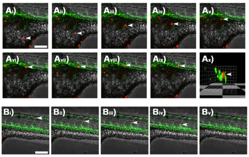
Single cell tracking by time-lapse confocal microscopy. Ai–ix) Confocal z-stack images taken at 15 minute intervals showing an individual DiI-labelled A375 melanoma cell (white arrows) migrating within the yolk sac of a casper zebrafish embryo and interacting with a GFP-tagged blood vessel. Ax) 3D-render of image Aix rotated to show the transverse section through the GFP-tagged blood vessel with DiI-labelled melanoma cell indicated by white arrows. Bi–v) Confocal z-stack images taken at 15 minute intervals showing an individual DiI-labelled melanoma cell (white arrows) within the GFP-tagged blood vessels of a casper zebrafish embryo. Scale bar = 150 μm. |

Representative confocal z-stack images of kdrl-GFP casper zebrafish embryos 72 hours after injection with human cancer cells. A) PC-3M-Pro4-mCherry prostate cancer cells injected into the duct of Cuvier form tumours in the caudal hematopoietic tissue of the zebrafish tail; Scale bar = 150 μm. B) Quantification of total mCherry fluorescence by prostate cancer cells after 1 and 3 days post injection; n=4, *p<0.01, 0.05 CI, paired t-test. Ci–ii) C8161 and Di–ii) WM164 melanoma cells (stained with Red DiI dye) injected alongside FluoSpheres (Blue) into the yolk sac survive and invade throughout the yolk sac; Scale bar = 500 μm. |