
- Title
-
Double maternal-effect: duplicated nucleoplasmin 2 genes, npm2a and npm2b, with essential but distinct functions are shared by fish and tetrapods
- Authors
- Cheung, C.T., Pasquier, J., Bouleau, A., Nguyen, T., Chesnel, F., Guiguen, Y., Bobe, J.
- Source
- Full text @ BMC Evol. Biol.

ZFIN is incorporating published figure images and captions as part of an ongoing project. Figures from some publications have not yet been curated, or are not available for display because of copyright restrictions. |
|
ZFIN is incorporating published figure images and captions as part of an ongoing project. Figures from some publications have not yet been curated, or are not available for display because of copyright restrictions. |
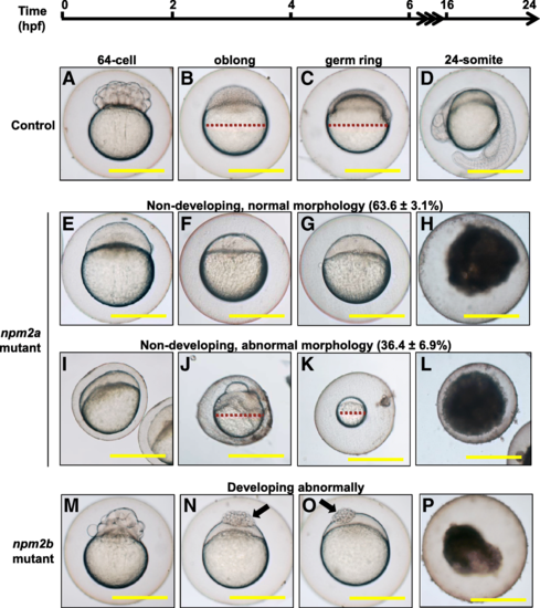
Effect of nucleoplasmin (npm) 2a and npm2b deficiencies on zebrafish embryogenesis. Representative images demonstrating development of fertilized eggs from crosses between control (a-d), npm2a (e-l), or npm2b (m-p) females and wildtype (WT) males from 2 to 24 h post-fertilization (hpf). In the control eggs, the embryos were at 64-cell (a), oblong (b), germ ring (c), and 24-somite (d) stages according to Kimmel et al. [23]. Eggs from npm2a mutant females were non-developing with a normal morphology (e-h) or with an abnormal morphology (i-l). Eggs from npm2b mutant females had a normal morphology albeit were developing abnormally (e-h). (a, e, i, m) = images taken at 2 hpf; (b, f, j, n) = images taken at 4 hpf; (c, g, k, o) = images taken at 6 hpf; (d, h, l, p) = images taken at 24 hpf. Scale bars denote 400 μm. Red dotted lines define the diameter of the embryo. Arrows demonstrate a partially cellularized blastodisc that was sitting atop an enlarged yolk syncytial layer |
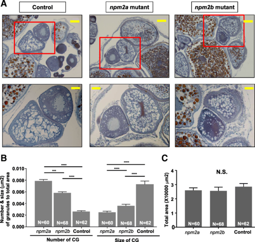
Histological analyses of the ovaries from nucleoplasmin (npm) 2a and npm2b mutant animals. a Ovary sections from age-matched npm2a, npm2b, and wildtype (WT) control females stained with Regaud’s hematoxylin. Top panels, 20X magnification; bars denote 90 μm; bottom panels show higher magnification images of the boxed areas in the top panels, 40X magnification; bars denote 45 μm. b Quantitation of the number and size (μm) of the cortical granules after adjusting to the total area of stage II follicles from ovaries from age-matched npm2a, npm2b, and control females. The N number denotes the total number of follicles counted for each group. c Total area of the stage II follicles from ovaries from age-matched npm2a, npm2b, and control females. N = 3 for all samples. N.S. = not significant, ***p < 0.001, ****p < 0.0001 by Mann-Whitney U-test PHENOTYPE:
|