
- Title
-
Characterizing sources of variability in zebrafish embryo screening protocols
- Authors
- Hamm, J.T., Ceger, P., Allen, D., Stout, M., Maull, E.A., Baker, G., Zmarowski, A., Padilla, S., Perkins, E., Planchart, A., Stedman, D., Tal, T., Tanguay, R.L., Volz, D.C., Wilbanks, M.S., Walker, N.J.
- Source
- Full text @ Altex

Zebrafish embryos in a 384-well tissue culture plate Photographs of transgenic fluorescent zebrafish embryos taken under a microscope using transmitted light in the top panel (A) and using fluorescence capture in the bottom panel (B). Images are captured simultaneously. One zebrafish embryo is immersed in 50 μl of embryo medium in each 3X3 mm well of the 384-well tissue culture plate. The embryos in this image were placed in the well at 5 hpf and the image was taken at 72 hpf. |
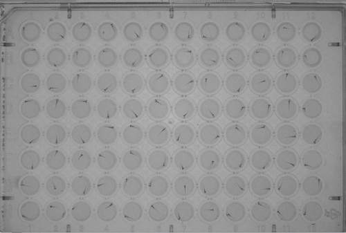
Zebrafish embryos in a 96-well tissue culture plate Photograph of zebrafish embryos in a 96-well tissue culture plate, taken under a microscope using transmitted light |