
- Title
-
Genome Editing Reveals Idiosyncrasy of CNGA2 Ion Channel-Directed Antibody Immunoreactivity Toward Oxytocin
- Authors
- Blechman, J., Anbalagan, S., Matthews, G.G., Levkowitz, G.
- Source
- Full text @ Front Cell Dev Biol
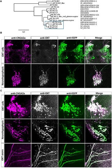
Immunoreactivity of anti-CNGA2a mAb in larval and adult zebrafish. (A) Phylogram of the CNG channels protein sequences. Comparison of the CNGA2 protein homologues from zebrafish, human, and mouse species is enclosed in the blue box. The scale bar indicates 30% amino acid residues substitution. (B,C) Confocal images showing representative labeling of the OXT perikarya, neuronal projections, and neurohypophyseal axonal termini with anti-CNGA2a mAb (magenta) and anti-OXT Ab (gray scale) in the context of the EGFP-positive OXT-ergic population in oxt:egfp reporter (green). Immunohistochemical analysis show colocalization of EGFP+, OXT+, and CNGA2a+ moieties in the cell bodies, axons, and nerve termini in the neurohypophysis of 6-day-old larva (n = 30/30) (B) and dissected brain and pituitary from 3-month-old adult zebrafish (n = 3/3) (C). Scale bars: 10 μm. |
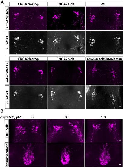
Anti-CNGA2a immunoreactivity is not affected following KO of cnga2a and/or cnga2b. (A) Immunostaining and confocal imaging of 6-day-old larva NPO of the wild type zebrafish (n = 30/30), cnga2a (n = 20/20), cnga2b (n = 16/16), and double cnga2a/2b (n = 6/6) zebrafish mutant variants with anti-CNGA (magenta) and anti-OXT (gray scale) antibodies. (B) Anti-CNGA2a mAb immunostaining and confocal imaging of the hypothalamus and neurohypophysis of 6-day-old larva injected with different concentrations of cnga2a missense morpholino oligonucleotide (n = 10/10). Scale bars: 10 μm. EXPRESSION / LABELING:
|

ZFIN is incorporating published figure images and captions as part of an ongoing project. Figures from some publications have not yet been curated, or are not available for display because of copyright restrictions. |
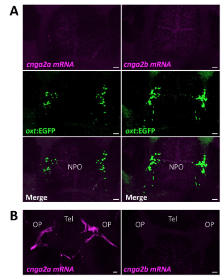
Expression of cnga2a/b mRNAs is not detected in zebrafish oxytocin neurons. (A) Confocal Z-stack images showing fluorescent in situ hybridization (FISH) of transgenic 6-dpf old Tg(oxt:EGFP) larvae using probes directed against mRNAs of cnga2a and cnga2b, followed by anti-EGFP staining. The NPO area with oxytocin cell bodies Tg(oxt:EGFP) are shown. No detectable expression of cnga2a and cnga2b is observed. Scale 10μm. (B) cnga2a but not cnga2b is expressed in olfactory placade of 6dpf old larvae using probes directed against mRNAs of cnga2a and cnga2b. Scale 10μm. EXPRESSION / LABELING:
|