
- Title
-
Hrg1 promotes heme-iron recycling during hemolysis in the zebrafish kidney
- Authors
- Zhang, J., Chambers, I., Yun, S., Phillips, J., Krause, M., Hamza, I.
- Source
- Full text @ PLoS Genet.
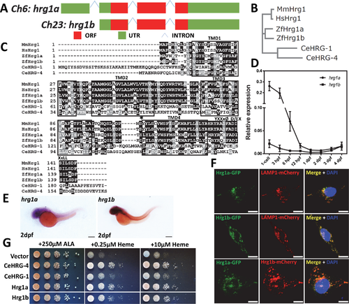
Hrg1 is duplicated in the zebrafish genome. (A) Two hrg1 homologs exist in zebrafish genome: hrg1a (slc48a1b) on chromosome 6 and hrg1b (slc48a1a) on chromosome 23. (B) Phylogenetic analysis of zebrafish Hrg1a and Hrg1b with CeHRG-1, CeHRG-4, MmHrg1, and Human Hrg1 (HsHrg1). Sequences were aligned using ClustalW and a phylogenetic tree was generated using the Neighbor-Joining method in MEGA5. (C) Multiple sequence alignment of zebrafish Hrg1a and Hrg1b with orthologs CeHRG-1, Cehrg-4, MmHrg1, and Human Hrg1 (HsHrg1). Asterisk, conserved histidine; black box, putative transmembrane domains; YXXxØ, C-terminal tyrosine sorting motif; D/EXXxLL, di-leucine sorting motif. (D) qRT-PCR of hrg1a and hrg1b in zebrafish embryos at different stages. 50 embryos before 24hpf were collected as one cohort. For embryos later than 24hpf, 30 were pooled as one cohort. 3 cohorts of each stage were harvested for total RNA extraction. Expression level was normalized to ß-actin. (E) Lateral view of hrg1a and hrg1b expression by WISH at 2dpf. Anterior is to the left. Scale bar: 200μm. (F) Confocal microscopy depicting subcellular localization of Hrg1a and Hrg1b with C-terminally tagged fluorescent proteins in transfected HEK-293 cells. Lamp1 is a marker for lysosomal compartments. Scale bar: 10 μm. (G) Yeast growth assay showing that zebrafish Hrg1a and Hrg1b are able to mediate heme transport in yeast. The hem1Δ strains transformed with empty vector pYes-DEST52, CeHRG-4, CeHRG-1, Hrg1a and Hrg1b were cultivated overnight and spotted in serial dilutions on SC plates supplemented with 250μM ALA and indicated concentrations of heme. EXPRESSION / LABELING:
|
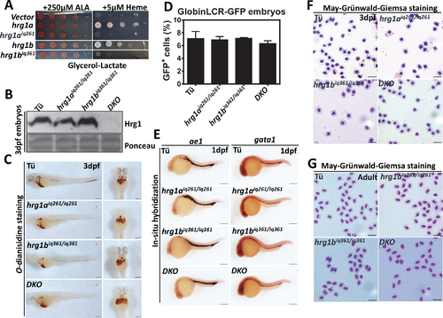
hrg1 DKO zebrafish survives to adulthood without overt hematological phenotypes. (A) Yeast growth assay showing that zebrafish Hrg1aiq261 and Hrg1biq361 alleles are incapable of mediating heme transport in yeast in contrast to WT forms of Hrg1a and Hrg1b. The hem1Δ strain transformed with empty vector pYes-DEST52, hrg1a, hrg1b, predicted hrg1aiq261 and hrg1biq361 were cultivated overnight and spotted in serial dilutions on SC plates supplemented with 250μM ALA and indicated concentration of heme. (B) Immunoblot of Hrg1 in membrane fractionation lysates from WT Tü, hrg1aiq261/iq261, hrg1biq361/iq361 and double mutant DKO. Each crude membrane fraction lysate was from pooled ~30 embryos. Each lane represents 100 μg of protein from membrane fractionation lysates. Ponceau is used as a loading control for membrane fraction. (C) O-dianisidine staining of RBCs in 3dpf embryos from Tü, hrg1aiq261/iq261, hrg1biq361/iq361 and DKO (n = 50 for each genotype). Scale bar: 200μm. (D) Quantitative percentages of GFP+ cells in Tü, hrg1aiq261/iq261, hrg1biq361/iq361 and DKO mutants in GlobinLCR-GFP transgenic background. Error bars indicate SEM of 3 independent experiments (two-way ANOVA, p >0.05). (E) Lateral view of ße1 and gata1 expression by WISH at 1dpf embryos from Tü, hrg1aiq261/iq261, hrg1biq361/iq361 and DKO. Anterior is to the left. N = 50 for each genotype. Scale bar: 200μm. (F) May-Grünwald-Giemsa staining of isolated peripheral RBCs at 3dpf embryos from Tü, hrg1aiq261/iq261, hrg1biq361/iq361 and hrg1aiq261/iq261; hrg1biq361/iq36 (N = 30). Scale bar: 20μm. (G) May-Grünwald-Giemsa staining of isolated peripheral RBCs from adult Tü, hrg1aiq261/iq261, hrg1biq361/iq361 and DKO. Scale bar: 20μm. EXPRESSION / LABELING:
PHENOTYPE:
|
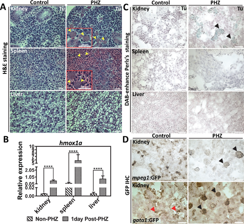
The zebrafish kidney is responsible for heme-iron recycling during EP. (A) H&E staining of kidney, spleen and liver sections from adult WT zebrafish with control (non-PHZ treated) and 1 day post PHZ-treatment samples. Cells undergoing erythrophagocytosis are indicated as yellow arrows. (B) qRT-PCR of hmox1a mRNA expression in the kidney, spleen and liver from control (non-PHZ treated) and PHZ treated adult zebrafish at 1day after treatment. Three adult zebrafish were pooled as one cohort, and three cohorts were repeated as biological triplicates. * p<0.05. (C) Perl’s Prussian blue iron staining on sections of kidney, spleen and liver from adult Tü WT zebrafish with control (non-PHZ treated) and 1 day post PHZ-treatment samples. Positive iron staining is indicated in kidney macrophages as showed by black arrows. (D) GFP IHC of kidney sections from adult transgenic zebrafish gata1:gfp and mpeg1:gfp with control (non-PHZ) and 1 day post PHZ-treatment. RBCs are indicated with red arrows and macrophages with black arrows. |
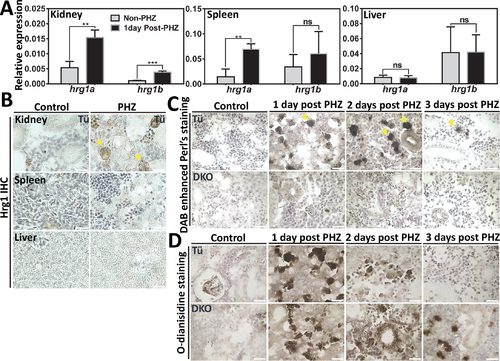
Defects of heme-iron recycling in kidney macrophages of Hrg1 DKO zebrafish. (A) qRT-PCR of hrg1a and hrg1b to quantify mRNA expression in the kidney, spleen and liver from control (non-PHZ treated) and 1-day post PHZ-treatment samples. Three adult zebrafish were pooled as one cohort, and three cohorts were repeated as biological triplicates. * p<0.05. (B) IHC staining of Hrg1 proteins in sections from the kidney, spleen and liver of adult zebrafish. (C) DAB-enhanced perl’s iron staining of kidney sections from Tü and hrg1 DKO zebrafish with control (non-PHZ), 1 day, 2 days and 3 days post PHZ-treatment. (D) O-dianisidine staining of kidney sections from Tü and hrg1 DKO zebrafish with control (non-PHZ), 1 day, 2 days and 3 days post PHZ-treatment. Scale bar: 20μm. |
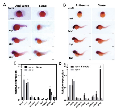
Expression of hrg1a and hrg1b in embryonic and adult zebrafish. (A-B) WISH of hrg1a and hrg1b expression in embryos at different stages. Anterior is to the left. Anti-sense probe is used to detect mRNA expression; sense probe is shown to indicate background staining. Scale bar: 200μm. (C-D) qRT-PCR on hrg1a and hrg1b in dissected adult zebrafish tissues from male and female. 3 male or female fish were dissected as one cohort, each gender had 3 cohorts as biological replicates. Expression level was normalized to ef1α. |
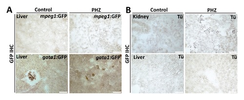
GFP IHC for zebrafish sections. (A) GFP IHC of liver sections from adult transgenic zebrafish gata1:gfp and mpeg1:gfp with control (non-PHZ) and 1 day post PHZ-treatment. (B) GFP IHC of kidney and liver sections from Tü WT zebrafish with control (non-PHZ) and 1 day post PHZ-treatment. |
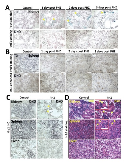
hrg1 DKO zebrafish undergoes normal erythrophagocytosis with defects in heme-iron recycling. (A) Perl’s Prussian blue iron staining of kidney sections from Tü and hrg1 DKO zebrafish with control (non-PHZ), 1 day, 2 days and 3 days post PHZ-treatment. Yellow arrows: macrophages. (B) DAB-enhanced perl’s iron staining of spleen sections from Tü and hrg1 DKO zebrafish with control (non-PHZ), 1 day, 2 days and 3 days post PHZ-treatment. (C) IHC staining of Hrg1 proteins in kidney, spleen and liver of adult hrg1 DKO zebrafish sections. (D) H&E staining of kidney, spleen and liver sections from hrg1 DKO zebrafish with control (non-PHZ) and 1-day post PHZ-treatment. Scale bar: 20μm. |

ZFIN is incorporating published figure images and captions as part of an ongoing project. Figures from some publications have not yet been curated, or are not available for display because of copyright restrictions. |