
- Title
-
Griseum centrale, a homologue of the periaqueductal gray in the lamprey.
- Authors
- Olson, I., Suryanarayana, S.M., Robertson, B., Grillner, S.
- Source
- Full text @ IBRO Rep

Identification of afferents to the griseum centrale. (A–C) Dual injections of Neurobiotin into the SNc (A) and biotin-dextran amine into the IPN (B). (B–C) Anterogradely labeled nerve fibers from IPN injection (red) in close proximity to retrogradely labeled cell bodies from SNc injection (green). (D–F) Injection of Neurobiotin into IPN (D) resulted in retrogradely labeled cells in the medial habenula (E-F; magenta). (G–H) Injection of Neurobiotin into griseum centrale (G) resulted in retrogradely labeled cells in the hypothalamus (H, arrow). (I) Pallial fibers and presumed terminals (red) in close apposition to cells in GC (green) retrogradely labeled from pretectum. Note that pallial fibers also innervated the contralateral GC. (J) Higher magnification of the photomicrograph in (I). (K) Neurobiotin injection into the lateral pallium (LPal). (L) Dextran injection into pretectum (PT). (M) GABA immunoreactive cells in the griseum centrale. All sections were counterstained with a fluorescent Nissl stain. IPN, interpeduncular nucleus; lHb, lateral habenula; M3, Müller cell 3; M5, the M5 nucleus of Schober; mHb, medial habenula nIII, oculomotor nerve. Scale bars = A, B, D, E, G, H, K, L, 250 μm; F, I 100 μm; C, I, J, M 50 μm. (For interpretation of the references to colour in this figure legend, the reader is referred to the web version of this article.) |
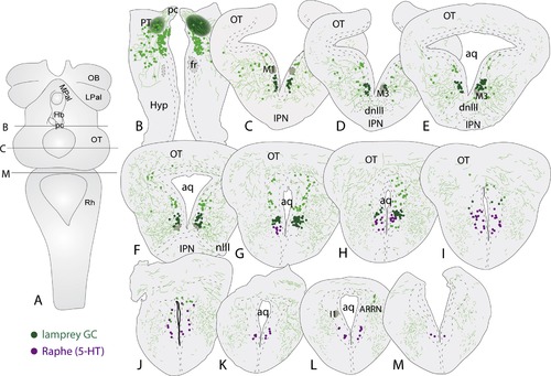
Location of the lamprey griseum centrale. (A) Dorsal view of the lamprey brain indicating the level of the injection site (B), as well as the most rostral (C) and the most caudal section (M). (B) The bilateral injection sites in the pretectum. (C–M) Schematic drawing illustrating the rostrocaudal extent of griseum centrale (GC; green) and its relation to the serotonergic raphe nucleus (magenta). Aq, aqueduct; ARRN, anterior rhombencephalic reticular nucleus; dnIII, dorsal nucleus of the oculomotor nerve; Hyp, hypothalamus; I1, I1 Müller cell; IPN, interpeduncular nucleus; nIII, the oculomotor nerve; OT, optic tectum; pc, posterior commissure; PT, pretectum. (For interpretation of the references to colour in this figure legend, the reader is referred to the web version of this article.) |